4K10

Crystal Structure of Leshmaniasis major Farnesyl diphosphate synthase in complex with 3-FLUORO-1-(2-HYDROXY-2,2-DIPHOSPHONOETHYL)PYRIDINIUM and Mg2+


Experimental Data Snapshot

  • Method: X-RAY DIFFRACTION
  • Resolution: 2.30 Å
  • R-Value Free: 0.276 
  • R-Value Work: 0.233 
  • R-Value Observed: 0.235 

wwPDB Validation   3D Report Full Report


Ligand Structure Quality Assessment 


This is version 1.2 of the entry. See complete history


Literature

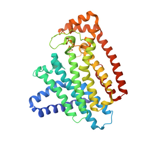
Structural and thermodynamic basis of the inhibition of Leishmania major farnesyl diphosphate synthase by nitrogen-containing bisphosphonates.

Aripirala, S.Gonzalez-Pacanowska, D.Oldfield, E.Kaiser, M.Amzel, L.M.Gabelli, S.B.

(2014) Acta Crystallogr D Biol Crystallogr 70: 802-810

  • DOI: https://doi.org/10.1107/S1399004713033221
  • Primary Citation of Related Structures:  
    4JZB, 4JZX, 4K10

  • PubMed Abstract: 

    Farnesyl diphosphate synthase (FPPS) is an essential enzyme involved in the biosynthesis of sterols (cholesterol in humans and ergosterol in yeasts, fungi and trypanosomatid parasites) as well as in protein prenylation. It is inhibited by bisphosphonates, a class of drugs used in humans to treat diverse bone-related diseases. The development of bisphosphonates as antiparasitic compounds targeting ergosterol biosynthesis has become an important route for therapeutic intervention. Here, the X-ray crystallographic structures of complexes of FPPS from Leishmania major (the causative agent of cutaneous leishmaniasis) with three bisphosphonates determined at resolutions of 1.8, 1.9 and 2.3 Å are reported. Two of the inhibitors, 1-(2-hydroxy-2,2-diphosphonoethyl)-3-phenylpyridinium (300B) and 3-butyl-1-(2,2-diphosphonoethyl)pyridinium (476A), co-crystallize with the homoallylic substrate isopentenyl diphosphate (IPP) and three Ca(2+) ions. A third inhibitor, 3-fluoro-1-(2-hydroxy-2,2-diphosphonoethyl)pyridinium (46I), was found to bind two Mg(2+) ions but not IPP. Calorimetric studies showed that binding of the inhibitors is entropically driven. Comparison of the structures of L. major FPPS (LmFPPS) and human FPPS provides new information for the design of bisphosphonates that will be more specific for inhibition of LmFPPS. The asymmetric structure of the LmFPPS-46I homodimer indicates that binding of the allylic substrate to both monomers of the dimer results in an asymmetric dimer with one open and one closed homoallylic site. It is proposed that IPP first binds to the open site, which then closes, opening the site on the other monomer, which closes after binding the second IPP, leading to the symmetric fully occupied FPPS dimer observed in other structures.


  • Organizational Affiliation

    Institute for Multiscale Modeling of Biological Interactions and Department of Biophysics and Biophysical Chemistry, Johns Hopkins University, 725 North Wolfe Street WBSB 605, Baltimore, MD 21210, USA.


Macromolecules
Find similar proteins by:  (by identity cutoff)  |  3D Structure
Entity ID: 1
MoleculeChains Sequence LengthOrganismDetailsImage
Farnesyl pyrophosphate synthaseA,
B,
C [auth D],
D [auth C]
362Leishmania majorMutation(s): 0 
Gene Names: FPPSLMJF_22_1360
EC: 2.5.1.1 (PDB Primary Data), 2.5.1.10 (PDB Primary Data)
UniProt
Find proteins for Q4QBL1 (Leishmania major)
Explore Q4QBL1 
Go to UniProtKB:  Q4QBL1
Entity Groups  
Sequence Clusters30% Identity50% Identity70% Identity90% Identity95% Identity100% Identity
UniProt GroupQ4QBL1
Sequence Annotations
Expand
  • Reference Sequence
Small Molecules
Ligands 6 Unique
IDChains Name / Formula / InChI Key2D Diagram3D Interactions
NI9
Query on NI9

Download Ideal Coordinates CCD File 
J [auth A],
P [auth B],
U [auth D],
Z [auth C]
3-FLUORO-1-(2-HYDROXY-2,2-DIPHOSPHONOETHYL)PYRIDINIUM
C7 H11 F N O7 P2
LSEAIOLITUIQJU-UHFFFAOYSA-O
MES
Query on MES

Download Ideal Coordinates CCD File 
Q [auth D]2-(N-MORPHOLINO)-ETHANESULFONIC ACID
C6 H13 N O4 S
SXGZJKUKBWWHRA-UHFFFAOYSA-N
PEG
Query on PEG

Download Ideal Coordinates CCD File 
K [auth A]DI(HYDROXYETHYL)ETHER
C4 H10 O3
MTHSVFCYNBDYFN-UHFFFAOYSA-N
ACT
Query on ACT

Download Ideal Coordinates CCD File 
M [auth B],
R [auth D]
ACETATE ION
C2 H3 O2
QTBSBXVTEAMEQO-UHFFFAOYSA-M
MG
Query on MG

Download Ideal Coordinates CCD File 
H [auth A]
I [auth A]
N [auth B]
O [auth B]
S [auth D]
H [auth A],
I [auth A],
N [auth B],
O [auth B],
S [auth D],
T [auth D],
X [auth C],
Y [auth C]
MAGNESIUM ION
Mg
JLVVSXFLKOJNIY-UHFFFAOYSA-N
NA
Query on NA

Download Ideal Coordinates CCD File 
E [auth A]
F [auth A]
G [auth A]
L [auth B]
V [auth C]
E [auth A],
F [auth A],
G [auth A],
L [auth B],
V [auth C],
W [auth C]
SODIUM ION
Na
FKNQFGJONOIPTF-UHFFFAOYSA-N
Binding Affinity Annotations 
IDSourceBinding Affinity
NI9 BindingDB:  4K10 Ki: 50 (nM) from 1 assay(s)
Binding MOAD:  4K10 Kd: 119.5 (nM) from 1 assay(s)
Experimental Data & Validation

Experimental Data

  • Method: X-RAY DIFFRACTION
  • Resolution: 2.30 Å
  • R-Value Free: 0.276 
  • R-Value Work: 0.233 
  • R-Value Observed: 0.235 
  • Space Group: P 21 21 21
Unit Cell:
Length ( Å )Angle ( ˚ )
a = 60.168α = 90
b = 143.743β = 90
c = 194.333γ = 90
Software Package:
Software NamePurpose
DENZOdata reduction
SCALEPACKdata scaling
REFMACrefinement
PDB_EXTRACTdata extraction
HKL-2000data scaling
AMoREphasing

Structure Validation

View Full Validation Report


Ligand Structure Quality Assessment 


Entry History 

Deposition Data

Revision History  (Full details and data files)

  • Version 1.0: 2014-04-02
    Type: Initial release
  • Version 1.1: 2017-11-15
    Changes: Refinement description
  • Version 1.2: 2024-02-28
    Changes: Data collection, Database references, Derived calculations