Crystal structures and biochemical studies of human lysophosphatidic acid phosphatase type 6
Li, J., Dong, Y., Lu, X., Wang, L., Peng, W., Zhang, X.C., Rao, Z.(2013) Protein Cell 4: 548-561
- PubMed: 23807634
- DOI: https://doi.org/10.1007/s13238-013-3031-z
- Primary Citation of Related Structures:
4JOB, 4JOC, 4JOD - PubMed Abstract:
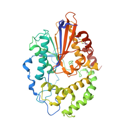
Lysophosphatidic acid (LPA) is an important bioactive phospholipid involved in cell signaling through Gprotein-coupled receptors pathways. It is also involved in balancing the lipid composition inside the cell, and modulates the function of lipid rafts as an intermediate in phospholipid metabolism. Because of its involvement in these important processes, LPA degradation needs to be regulated as precisely as its production. Lysophosphatidic acid phosphatase type 6 (ACP6) is an LPA-specific acid phosphatase that hydrolyzes LPA to monoacylglycerol (MAG) and phosphate. Here, we report three crystal structures of human ACP6 in complex with malonate, L-(+)-tartrate and tris, respectively. Our analyses revealed that ACP6 possesses a highly conserved Rossmann-foldlike body domain as well as a less conserved cap domain. The vast hydrophobic substrate-binding pocket, which is located between those two domains, is suitable for accommodating LPA, and its shape is different from that of other histidine acid phosphatases, a fact that is consistent with the observed difference in substrate preferences. Our analysis of the binding of three molecules in the active site reveals the involvement of six conserved and crucial residues in binding of the LPA phosphate group and its catalysis. The structure also indicates a water-supplying channel for substrate hydrolysis. Our structural data are consistent with the fact that the enzyme is active as a monomer. In combination with additional mutagenesis and enzyme activity studies, our structural data provide important insights into substrate recognition and the mechanism for catalytic activity of ACP6.
Organizational Affiliation:
National Laboratory of Biomacromolecules, Institute of Biophysics, Chinese Academy of Sciences, Beijing 100101, China.