4JEA

Crystal structure of an engineered Zn-RIDC1 construct with four interfacial disulfide bonds


Experimental Data Snapshot

  • Method: X-RAY DIFFRACTION
  • Resolution: 1.22 Å
  • R-Value Free: 0.179 
  • R-Value Work: 0.131 
  • R-Value Observed: 0.134 

wwPDB Validation   3D Report Full Report


Ligand Structure Quality Assessment 


This is version 1.2 of the entry. See complete history


Literature

In Vitro and Cellular Self-Assembly of a Zn-Binding Protein Cryptand via Templated Disulfide Bonds.

Medina-Morales, A.Perez, A.Brodin, J.D.Tezcan, F.A.

(2013) J Am Chem Soc 135: 12013-12022

  • DOI: https://doi.org/10.1021/ja405318d
  • Primary Citation of Related Structures:  
    4JE9, 4JEA, 4JEB

  • PubMed Abstract: 

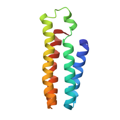
    Simultaneously strong and reversible through redox chemistry, disulfide bonds play a unique and often irreplaceable role in the formation of biological and synthetic assemblies. In an approach inspired by supramolecular chemistry, we report here that engineered noncovalent interactions on the surface of a monomeric protein can template its assembly into a unique cryptand-like protein complex ((C81/C96)RIDC14) by guiding the selective formation of multiple disulfide bonds across different interfaces. Owing to its highly interconnected framework, (C81/C96)RIDC14 is well preorganized for metal coordination in its interior, can support a large internal cavity surrounding the metal sites, and can withstand significant alterations in inner-sphere metal coordination. (C81/C96)RIDC14 self-assembles with high fidelity and yield in the periplasmic space of E. coli cells, where it can successfully compete for Zn(II) binding.


  • Organizational Affiliation

    Department of Chemistry and Biochemistry, University of California, San Diego, La Jolla, California 92093-0356, USA.


Macromolecules
Find similar proteins by:  (by identity cutoff)  |  3D Structure
Entity ID: 1
MoleculeChains Sequence LengthOrganismDetailsImage
soluble cytochrome b562
A, B, C, D
106Escherichia coliMutation(s): 13 
Gene Names: cybC
UniProt
Find proteins for P0ABE7 (Escherichia coli)
Explore P0ABE7 
Go to UniProtKB:  P0ABE7
Entity Groups  
Sequence Clusters30% Identity50% Identity70% Identity90% Identity95% Identity100% Identity
UniProt GroupP0ABE7
Sequence Annotations
Expand
  • Reference Sequence
Small Molecules
Ligands 3 Unique
IDChains Name / Formula / InChI Key2D Diagram3D Interactions
HEM
Query on HEM

Download Ideal Coordinates CCD File 
E [auth A],
H [auth B],
K [auth C],
O [auth D]
PROTOPORPHYRIN IX CONTAINING FE
C34 H32 Fe N4 O4
KABFMIBPWCXCRK-RGGAHWMASA-L
SO4
Query on SO4

Download Ideal Coordinates CCD File 
G [auth A]
J [auth B]
M [auth C]
N [auth C]
Q [auth D]
G [auth A],
J [auth B],
M [auth C],
N [auth C],
Q [auth D],
R [auth D],
S [auth D]
SULFATE ION
O4 S
QAOWNCQODCNURD-UHFFFAOYSA-L
ZN
Query on ZN

Download Ideal Coordinates CCD File 
F [auth A],
I [auth B],
L [auth C],
P [auth D]
ZINC ION
Zn
PTFCDOFLOPIGGS-UHFFFAOYSA-N
Experimental Data & Validation

Experimental Data

  • Method: X-RAY DIFFRACTION
  • Resolution: 1.22 Å
  • R-Value Free: 0.179 
  • R-Value Work: 0.131 
  • R-Value Observed: 0.134 
  • Space Group: P 1 21 1
Unit Cell:
Length ( Å )Angle ( ˚ )
a = 48.85α = 90
b = 64.788β = 98.18
c = 74.177γ = 90
Software Package:
Software NamePurpose
SCALAdata scaling
REFMACrefinement
PDB_EXTRACTdata extraction
MOSFLMdata reduction
PHASERphasing

Structure Validation

View Full Validation Report


Ligand Structure Quality Assessment 


Entry History 

Deposition Data

Revision History  (Full details and data files)

  • Version 1.0: 2013-08-21
    Type: Initial release
  • Version 1.1: 2013-08-28
    Changes: Database references
  • Version 1.2: 2023-09-20
    Changes: Data collection, Database references, Derived calculations, Refinement description