4J7H

Crystal structure of EvaA, a 2,3-dehydratase in complex with dTDP-benzene and dTDP-rhamnose


Experimental Data Snapshot

  • Method: X-RAY DIFFRACTION
  • Resolution: 1.69 Å
  • R-Value Free: 0.191 
  • R-Value Work: 0.165 
  • R-Value Observed: 0.166 

wwPDB Validation   3D Report Full Report


Ligand Structure Quality Assessment 


This is version 1.2 of the entry. See complete history


Literature

Structure of EvaA: A Paradigm for Sugar 2,3-Dehydratases.

Kubiak, R.L.Thoden, J.B.Holden, H.M.

(2013) Biochemistry 52: 2078-2088

  • DOI: https://doi.org/10.1021/bi400176n
  • Primary Citation of Related Structures:  
    4J7G, 4J7H

  • PubMed Abstract: 

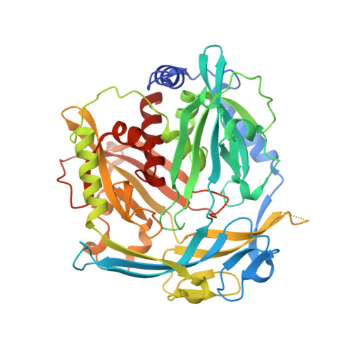
    Unusual deoxysugars found appended to natural products often provide or enhance the pharmacokinetic activities of the parent compound. The preferred carbohydrate donors for the biosynthesis of such glycosylated natural products are the dTDP-linked sugars. Many of the biologically relevant dTDP-deoxysugars are constructed around the 2,6-dideoxyhexoses or the 2,3(4),6-trideoxyhexoses. A key step in the biosynthesis of these sugars is the removal of the hexose C-2' hydroxyl group and the oxidation of the C-3' hydroxyl group to a carbonyl moiety. Enzymes that catalyze these reactions are referred to as 2,3-dehydratases and have been, for the most part, largely uncharacterized. Here we report the first structural analysis of a sugar 2,3-dehydratase. For this investigation, the enzyme, EvaA, was cloned from Amycolatopsis orientalis, and the structure was solved and refined to a nominal resolution of 1.7 Å. On the basis of the resulting model, it is clear that EvaA belongs to the large Nudix hydrolase superfamily and is most similar to GDP-mannose hydrolase. Each subunit of the EvaA dimer folds into two domains that clearly arose via gene duplication. Two dTDP-sugar binding pockets, A and B, are present in each EvaA subunit. On the basis of site-directed mutagenesis experiments and activity assays, it appears that pocket A functions as the active site and pocket B is simply a remnant left behind from the gene duplication event. As 2,3-dehydration is crucial for the biosynthesis of many unusual deoxysugars, this investigation provides key structural insight into this widely conserved reaction.


  • Organizational Affiliation

    Department of Biochemistry, University of Wisconsin , Madison, Wisconsin 53706, United States.


Macromolecules
Find similar proteins by:  (by identity cutoff)  |  3D Structure
Entity ID: 1
MoleculeChains Sequence LengthOrganismDetailsImage
EvaA 2,3-dehydratase
A, B
471Amycolatopsis orientalisMutation(s): 1 
Gene Names: EvaA
UniProt
Find proteins for O52793 (Amycolatopsis orientalis)
Explore O52793 
Go to UniProtKB:  O52793
Entity Groups  
Sequence Clusters30% Identity50% Identity70% Identity90% Identity95% Identity100% Identity
UniProt GroupO52793
Sequence Annotations
Expand
  • Reference Sequence
Small Molecules
Ligands 3 Unique
IDChains Name / Formula / InChI Key2D Diagram3D Interactions
TRH
Query on TRH

Download Ideal Coordinates CCD File 
D [auth A],
J [auth B]
2'-DEOXY-THYMIDINE-BETA-L-RHAMNOSE
C16 H26 N2 O15 P2
ZOSQFDVXNQFKBY-CGAXJHMRSA-N
TLO
Query on TLO

Download Ideal Coordinates CCD File 
C [auth A],
I [auth B]
5'-O-[(S)-hydroxy{[(S)-hydroxy(phenoxy)phosphoryl]oxy}phosphoryl]thymidine
C16 H20 N2 O11 P2
BANJVYKYIQPYKP-BFHYXJOUSA-N
EDO
Query on EDO

Download Ideal Coordinates CCD File 
E [auth A]
F [auth A]
G [auth A]
H [auth A]
K [auth B]
E [auth A],
F [auth A],
G [auth A],
H [auth A],
K [auth B],
L [auth B]
1,2-ETHANEDIOL
C2 H6 O2
LYCAIKOWRPUZTN-UHFFFAOYSA-N
Experimental Data & Validation

Experimental Data

  • Method: X-RAY DIFFRACTION
  • Resolution: 1.69 Å
  • R-Value Free: 0.191 
  • R-Value Work: 0.165 
  • R-Value Observed: 0.166 
  • Space Group: P 21 21 2
Unit Cell:
Length ( Å )Angle ( ˚ )
a = 104.681α = 90
b = 108.635β = 90
c = 110.133γ = 90
Software Package:
Software NamePurpose
HKL-3000data collection
PHASERphasing
REFMACrefinement
HKL-3000data reduction
HKL-3000data scaling

Structure Validation

View Full Validation Report


Ligand Structure Quality Assessment 


Entry History 

Deposition Data

Revision History  (Full details and data files)

  • Version 1.0: 2013-05-22
    Type: Initial release
  • Version 1.1: 2013-11-20
    Changes: Database references
  • Version 1.2: 2013-12-25
    Changes: Database references