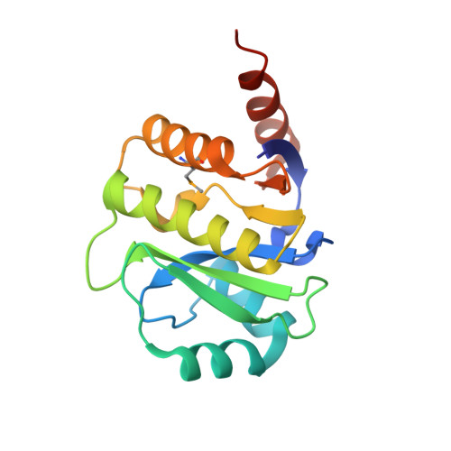
Crystal structure of the improved variant of the evolved serine hydrolase, OSH55.4_H1.2, bond with sulfate ion in the active site, Northeast Structural Genomics Consortium (NESG) Target OR301
Kuzin, A.P., Lew, S., Rajagopalan, S., Maglaqui, M., Xiao, R., Lee, D., Everett, J.K., Acton, T.B., Baker, D., Montelione, G.T., Tong, L., Hunt, J.F.To be published.