4ISA

Crystal Structure of the Escherichia coli LpxC/BB-78485 complex


Experimental Data Snapshot

  • Method: X-RAY DIFFRACTION
  • Resolution: 1.80 Å
  • R-Value Free: 0.211 
  • R-Value Work: 0.183 
  • R-Value Observed: 0.185 

wwPDB Validation   3D Report Full Report


Ligand Structure Quality Assessment 


This is version 1.3 of the entry. See complete history


Literature

Structural Basis of the Promiscuous Inhibitor Susceptibility of Escherichia coli LpxC.

Lee, C.J.Liang, X.Gopalaswamy, R.Najeeb, J.Ark, E.D.Toone, E.J.Zhou, P.

(2014) ACS Chem Biol 9: 237-246

  • DOI: https://doi.org/10.1021/cb400067g
  • Primary Citation of Related Structures:  
    4IS9, 4ISA, 4MQY

  • PubMed Abstract: 

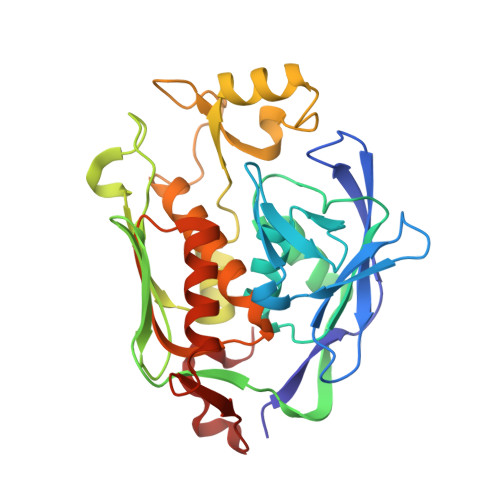
    The LpxC enzyme in the lipid A biosynthetic pathway is one of the most promising and clinically unexploited antibiotic targets for treatment of multidrug-resistant Gram-negative infections. Progress in medicinal chemistry has led to the discovery of potent LpxC inhibitors with a variety of chemical scaffolds and distinct antibiotic profiles. The vast majority of these compounds, including the nanomolar inhibitors L-161,240 and BB-78485, are highly effective in suppressing the activity of Escherichia coli LpxC (EcLpxC) but not divergent orthologs such as Pseudomonas aeruginosa LpxC (PaLpxC) in vitro. The molecular basis for such promiscuous inhibition of EcLpxC has remained poorly understood. Here, we report the crystal structure of EcLpxC bound to L-161,240, providing the first molecular insight into L-161,240 inhibition. Additionally, structural analysis of the EcLpxC/L-161,240 complex together with the EcLpxC/BB-78485 complex reveals an unexpected backbone flipping of the Insert I βa-βb loop in EcLpxC in comparison with previously reported crystal structures of EcLpxC complexes with l-threonyl-hydroxamate-based broad-spectrum inhibitors. Such a conformational switch, which has only been observed in EcLpxC but not in divergent orthologs such as PaLpxC, results in expansion of the active site of EcLpxC, enabling it to accommodate LpxC inhibitors with a variety of head groups, including compounds containing single (R- or S-enantiomers) or double substitutions at the neighboring Cα atom of the hydroxamate warhead group. These results highlight the importance of understanding inherent conformational plasticity of target proteins in lead optimization.


  • Organizational Affiliation

    Department of Biochemistry, Duke University Medical Center , Durham, North Carolina 27710, United States.


Macromolecules
Find similar proteins by:  (by identity cutoff)  |  3D Structure
Entity ID: 1
MoleculeChains Sequence LengthOrganismDetailsImage
UDP-3-O-[3-hydroxymyristoyl] N-acetylglucosamine deacetylase300Escherichia coli IHE3034Mutation(s): 0 
Gene Names: lpxCECOK1_0097
EC: 3.5.1
Entity Groups  
Sequence Clusters30% Identity50% Identity70% Identity90% Identity95% Identity100% Identity
Sequence Annotations
Expand
  • Reference Sequence
Small Molecules
Ligands 4 Unique
IDChains Name / Formula / InChI Key2D Diagram3D Interactions
GVR
Query on GVR

Download Ideal Coordinates CCD File 
C [auth A],
D [auth A]
(2R)-N-hydroxy-3-naphthalen-2-yl-2-[(naphthalen-2-ylsulfonyl)amino]propanamide
C23 H20 N2 O4 S
MMOUXLMPQFMDRD-JOCHJYFZSA-N
D1D
Query on D1D

Download Ideal Coordinates CCD File 
V [auth A](4S,5S)-1,2-DITHIANE-4,5-DIOL
C4 H8 O2 S2
YPGMOWHXEQDBBV-QWWZWVQMSA-N
ZN
Query on ZN

Download Ideal Coordinates CCD File 
B [auth A]ZINC ION
Zn
PTFCDOFLOPIGGS-UHFFFAOYSA-N
FMT
Query on FMT

Download Ideal Coordinates CCD File 
E [auth A]
F [auth A]
G [auth A]
H [auth A]
I [auth A]
E [auth A],
F [auth A],
G [auth A],
H [auth A],
I [auth A],
J [auth A],
K [auth A],
L [auth A],
M [auth A],
N [auth A],
O [auth A],
P [auth A],
Q [auth A],
R [auth A],
S [auth A],
T [auth A],
U [auth A]
FORMIC ACID
C H2 O2
BDAGIHXWWSANSR-UHFFFAOYSA-N
Binding Affinity Annotations 
IDSourceBinding Affinity
GVR BindingDB:  4ISA Ki: min: 0.07, max: 20 (nM) from 3 assay(s)
Experimental Data & Validation

Experimental Data

  • Method: X-RAY DIFFRACTION
  • Resolution: 1.80 Å
  • R-Value Free: 0.211 
  • R-Value Work: 0.183 
  • R-Value Observed: 0.185 
  • Space Group: P 61
Unit Cell:
Length ( Å )Angle ( ˚ )
a = 106.788α = 90
b = 106.788β = 90
c = 52.474γ = 120
Software Package:
Software NamePurpose
DENZOdata reduction
SCALEPACKdata scaling
PHENIXrefinement
PDB_EXTRACTdata extraction
StructureStudiodata collection

Structure Validation

View Full Validation Report


Ligand Structure Quality Assessment 


Entry History 

Deposition Data

Revision History  (Full details and data files)

  • Version 1.0: 2013-10-30
    Type: Initial release
  • Version 1.1: 2014-02-12
    Changes: Database references
  • Version 1.2: 2017-11-15
    Changes: Refinement description
  • Version 1.3: 2024-02-28
    Changes: Data collection, Database references, Derived calculations