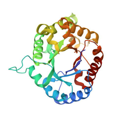
4IOT

High-resolution Structure of Triosephosphate isomerase from E. coli


Experimental Data Snapshot

  • Method: X-RAY DIFFRACTION
  • Resolution: 1.85 Å
  • R-Value Free: 0.206 
  • R-Value Work: 0.172 
  • R-Value Observed: 0.174 

wwPDB Validation   3D Report Full Report


This is version 1.2 of the entry. See complete history


Literature

Triosephosphate isomerase is a common crystallization contaminant of soluble His-tagged proteins produced in Escherichia coli.

Kozlov, G.Vinaik, R.Gehring, K.

(2013) Acta Crystallogr Sect F Struct Biol Cryst Commun 69: 499-502

  • DOI: https://doi.org/10.1107/S1744309113010841
  • Primary Citation of Related Structures:  
    4IOT

  • PubMed Abstract: 

    Attempts to crystallize several mammalian proteins overexpressed in Escherichia coli revealed a common contaminant, triosephosphate isomerase, a protein involved in glucose metabolism. Even with triosephosphate isomerase present in very small amounts, similarly shaped crystals appeared in the crystallization drops in a number of polyethylene glycol-containing conditions. All of the target proteins were His-tagged and their purification involved immobilized metal-affinity chromatography (IMAC), a step that was likely to lead to triosephosphate isomerase contamination. Analysis of the triosephosphate isomerase crystals led to the structure of E. coli triosephosphate isomerase at 1.85 Å resolution, which is a significant improvement over the previous structure.


  • Organizational Affiliation

    Department of Biochemistry, Groupe de Recherche axé sur la Structure des Protéines, McGill University, 3649 Promenade Sir William Osler, Montréal, Québec H3G 0B1, Canada.


Macromolecules
Find similar proteins by:  (by identity cutoff)  |  3D Structure
Entity ID: 1
MoleculeChains Sequence LengthOrganismDetailsImage
Triosephosphate isomerase
A, B
255Escherichia coli str. K-12 substr. DH10BMutation(s): 0 
EC: 5.3.1.1
UniProt
Find proteins for B1XB85 (Escherichia coli (strain K12 / DH10B))
Explore B1XB85 
Go to UniProtKB:  B1XB85
Entity Groups  
Sequence Clusters30% Identity50% Identity70% Identity90% Identity95% Identity100% Identity
UniProt GroupB1XB85
Sequence Annotations
Expand
  • Reference Sequence
Small Molecules
Ligands 1 Unique
IDChains Name / Formula / InChI Key2D Diagram3D Interactions
SO4
Query on SO4

Download Ideal Coordinates CCD File 
C [auth A]SULFATE ION
O4 S
QAOWNCQODCNURD-UHFFFAOYSA-L
Experimental Data & Validation

Experimental Data

  • Method: X-RAY DIFFRACTION
  • Resolution: 1.85 Å
  • R-Value Free: 0.206 
  • R-Value Work: 0.172 
  • R-Value Observed: 0.174 
  • Space Group: P 21 21 21
Unit Cell:
Length ( Å )Angle ( ˚ )
a = 46.47α = 90
b = 67.452β = 90
c = 150.354γ = 90
Software Package:
Software NamePurpose
ADSCdata collection
PHASERphasing
REFMACrefinement
HKL-2000data reduction
HKL-2000data scaling

Structure Validation

View Full Validation Report


Entry History 

Deposition Data

Revision History  (Full details and data files)

  • Version 1.0: 2013-01-23
    Type: Initial release
  • Version 1.1: 2013-06-05
    Changes: Database references
  • Version 1.2: 2023-09-20
    Changes: Data collection, Database references, Derived calculations, Refinement description