4IKR

Crystal structure of Type 1 human methionine aminopeptidase in complex with 2-(4-(5-chloro-6-methyl-2-(pyridin-2-yl)pyrimidin-4-yl)piperazin-1-yl)ethanol


Experimental Data Snapshot

  • Method: X-RAY DIFFRACTION
  • Resolution: 1.78 Å
  • R-Value Free: 0.211 
  • R-Value Work: 0.180 
  • R-Value Observed: 0.182 

wwPDB Validation   3D Report Full Report


Ligand Structure Quality Assessment 


This is version 1.2 of the entry. See complete history


Literature

Identification, Biochemical and Structural Evaluation of Species-Specific Inhibitors against Type I Methionine Aminopeptidases

Kishor, C.Arya, T.Reddi, R.Chen, X.Saddanapu, V.Marapaka, A.K.Gumpena, R.Ma, D.Liu, J.O.Addlagatta, A.

(2013) J Med Chem 56: 5295-5305

  • DOI: https://doi.org/10.1021/jm400395p
  • Primary Citation of Related Structures:  
    4IKR, 4IKS, 4IKT, 4IKU

  • PubMed Abstract: 

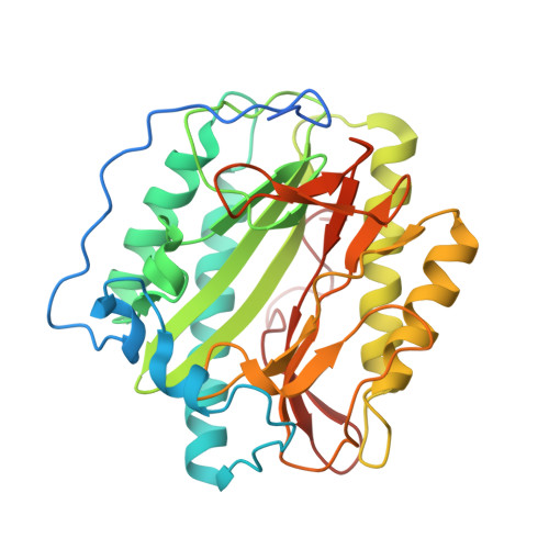
    Methionine aminopeptidases (MetAPs) are essential enzymes that make them good drug targets in cancer and microbial infections. MetAPs remove the initiator methionine from newly synthesized peptides in every living cell. MetAPs are broadly divided into type I and type II classes. Both prokaryotes and eukaryotes contain type I MetAPs, while eukaryotes have additional type II MetAP enzyme. Although several inhibitors have been reported against type I enzymes, subclass specificity is scarce. Here, using the fine differences in the entrance of the active sites of MetAPs from Mycobacterium tuberculosis , Enterococcus faecalis , and human, three hotspots have been identified and pyridinylpyrimidine-based molecules were selected from a commercial source to target these hotspots. In the biochemical evaluation, many of the 38 compounds displayed differential behavior against these three enzymes. Crystal structures of four selected inhibitors in complex with human MetAP1b and molecular modeling studies provided the basis for the binding specificity.


  • Organizational Affiliation

    Center for Chemical Biology, CSIR-Indian Institute of Chemical Technology , Tarnaka, Hyderabad AP-500 007, India.


Macromolecules
Find similar proteins by:  (by identity cutoff)  |  3D Structure
Entity ID: 1
MoleculeChains Sequence LengthOrganismDetailsImage
Methionine aminopeptidase 1329Homo sapiensMutation(s): 0 
Gene Names: METAP1
EC: 3.4.11.18
UniProt & NIH Common Fund Data Resources
Find proteins for P53582 (Homo sapiens)
Explore P53582 
Go to UniProtKB:  P53582
PHAROS:  P53582
GTEx:  ENSG00000164024 
Entity Groups  
Sequence Clusters30% Identity50% Identity70% Identity90% Identity95% Identity100% Identity
UniProt GroupP53582
Sequence Annotations
Expand
  • Reference Sequence
Small Molecules
Binding Affinity Annotations 
IDSourceBinding Affinity
PVP Binding MOAD:  4IKR IC50: 590 (nM) from 1 assay(s)
PDBBind:  4IKR IC50: 590 (nM) from 1 assay(s)
BindingDB:  4IKR IC50: 1100 (nM) from 1 assay(s)
Experimental Data & Validation

Experimental Data

  • Method: X-RAY DIFFRACTION
  • Resolution: 1.78 Å
  • R-Value Free: 0.211 
  • R-Value Work: 0.180 
  • R-Value Observed: 0.182 
  • Space Group: P 1 21 1
Unit Cell:
Length ( Å )Angle ( ˚ )
a = 47.345α = 90
b = 77.423β = 91.03
c = 48.023γ = 90
Software Package:
Software NamePurpose
HKL-3000data collection
MOLREPphasing
REFMACrefinement
HKL-3000data reduction
HKL-3000data scaling

Structure Validation

View Full Validation Report


Ligand Structure Quality Assessment 


Entry History 

Deposition Data

Revision History  (Full details and data files)

  • Version 1.0: 2013-12-11
    Type: Initial release
  • Version 1.1: 2019-11-20
    Changes: Database references, Derived calculations
  • Version 1.2: 2024-03-20
    Changes: Data collection, Database references, Derived calculations