4I3V

Structure of phosphonoacetaldehyde dehydrogenase in complex with phosphonoacetaldehyde and cofactor NAD+


Experimental Data Snapshot

  • Method: X-RAY DIFFRACTION
  • Resolution: 2.00 Å
  • R-Value Free: 0.230 
  • R-Value Work: 0.182 
  • R-Value Observed: 0.185 

wwPDB Validation   3D Report Full Report


Ligand Structure Quality Assessment 


This is version 1.2 of the entry. See complete history


Literature

Structure and function of phosphonoacetaldehyde dehydrogenase: the missing link in phosphonoacetate formation.

Agarwal, V.Peck, S.C.Chen, J.H.Borisova, S.A.Chekan, J.R.van der Donk, W.A.Nair, S.K.

(2014) Chem Biol 21: 125-135

  • DOI: https://doi.org/10.1016/j.chembiol.2013.11.006
  • Primary Citation of Related Structures:  
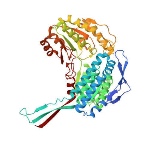
    4I3T, 4I3U, 4I3V, 4I3W, 4I3X

  • PubMed Abstract: 

    Phosphonates (C-PO₃²⁻) have applications as antibiotics, herbicides, and detergents. In some environments, these molecules represent the predominant source of phosphorus, and several microbes have evolved dedicated enzymatic machineries for phosphonate degradation. For example, most common naturally occurring phosphonates can be catabolized to either phosphonoacetaldehyde or phosphonoacetate, which can then be hydrolyzed to generate inorganic phosphate and acetaldehyde or acetate, respectively. The phosphonoacetaldehyde oxidase gene (phnY) links these two hydrolytic processes and provides a previously unknown catabolic mechanism for phosphonoacetate production in the microbial metabolome. Here, we present biochemical characterization of PhnY and high-resolution crystal structures of the apo state, as well as complexes with substrate, cofactor, and product. Kinetic analysis of active site mutants demonstrates how a highly conserved aldehyde dehydrogenase active site has been modified in nature to generate activity with a phosphonate substrate.


  • Organizational Affiliation

    Center for Biophysics and Computational Biology, University of Illinois at Urbana-Champaign, Urbana, IL 61801, USA; Institute for Genomic Biology, University of Illinois at Urbana-Champaign, Urbana, IL 61801, USA.


Macromolecules
Find similar proteins by:  (by identity cutoff)  |  3D Structure
Entity ID: 1
MoleculeChains Sequence LengthOrganismDetailsImage
Aldehyde dehydrogenase (NAD+)
A, B, C, D, E
A, B, C, D, E, F, G, H
488Sinorhizobium meliloti 1021Mutation(s): 0 
Gene Names: phnYRB0979SM_b21539
EC: 1.2.1.3
UniProt
Find proteins for Q92UV7 (Rhizobium meliloti (strain 1021))
Explore Q92UV7 
Go to UniProtKB:  Q92UV7
Entity Groups  
Sequence Clusters30% Identity50% Identity70% Identity90% Identity95% Identity100% Identity
UniProt GroupQ92UV7
Sequence Annotations
Expand
  • Reference Sequence
Small Molecules
Ligands 2 Unique
IDChains Name / Formula / InChI Key2D Diagram3D Interactions
NAD
Query on NAD

Download Ideal Coordinates CCD File 
I [auth A]
K [auth B]
M [auth C]
O [auth D]
Q [auth E]
I [auth A],
K [auth B],
M [auth C],
O [auth D],
Q [auth E],
S [auth F],
U [auth G]
NICOTINAMIDE-ADENINE-DINUCLEOTIDE
C21 H27 N7 O14 P2
BAWFJGJZGIEFAR-NNYOXOHSSA-N
POA
Query on POA

Download Ideal Coordinates CCD File 
J [auth A]
L [auth B]
N [auth C]
P [auth D]
R [auth E]
J [auth A],
L [auth B],
N [auth C],
P [auth D],
R [auth E],
T [auth F],
V [auth G],
W [auth H]
PHOSPHONOACETALDEHYDE
C2 H5 O4 P
YEMKIGUKNDOZEG-UHFFFAOYSA-N
Experimental Data & Validation

Experimental Data

  • Method: X-RAY DIFFRACTION
  • Resolution: 2.00 Å
  • R-Value Free: 0.230 
  • R-Value Work: 0.182 
  • R-Value Observed: 0.185 
  • Space Group: P 1 21 1
Unit Cell:
Length ( Å )Angle ( ˚ )
a = 94.744α = 90
b = 172.128β = 107.75
c = 140.056γ = 90
Software Package:
Software NamePurpose
DENZOdata reduction
SCALEPACKdata scaling
REFMACrefinement
PDB_EXTRACTdata extraction
PHENIXrefinement

Structure Validation

View Full Validation Report


Ligand Structure Quality Assessment 


Entry History 

Deposition Data

Revision History  (Full details and data files)

  • Version 1.0: 2013-11-27
    Type: Initial release
  • Version 1.1: 2014-02-05
    Changes: Database references
  • Version 1.2: 2017-11-15
    Changes: Refinement description