Redefining the Role of the Quaternary Shift in Bacillus stearothermophilus Phosphofructokinase.
Mosser, R., Reddy, M.C., Bruning, J.B., Sacchettini, J.C., Reinhart, G.D.(2013) Biochemistry 52: 5421-5429
- PubMed: 23859543
- DOI: https://doi.org/10.1021/bi4002503
- Primary Citation of Related Structures:
4I36, 4I4I, 4I7E - PubMed Abstract:
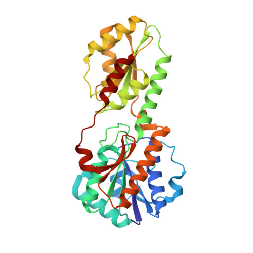
Bacillus stearothermophilus phosphofructokinase (BsPFK) is a homotetramer that is allosterically inhibited by phosphoenolpyruvate (PEP), which binds along one dimer-dimer interface. The substrate, fructose 6-phosphate (Fru-6-P), binds along the other dimer-dimer interface. Evans et al. observed that the structure with inhibitor (phosphoglycolate) bound, compared to the structure of wild-type BsPFK with substrate and activator bound, exhibits a 7° rotation about the substrate-binding interface, termed the quaternary shift [Schirmer, T., and Evans, P. R. (1990) Nature 343, 140-145]. We report that the variant D12A BsPFK exhibits a 100-fold increase in its binding affinity for PEP, a 50-fold decrease in its binding affinity for Fru-6-P, but an inhibitory coupling comparable to that of the wild type. Crystal structures of the apo and PEP-bound forms of D12A BsPFK have been determined (Protein Data Bank entries 4I36 and 4I7E , respectively), and both indicate a shifted structure similar to the inhibitor-bound structure of the wild type. D12 does not directly bind to either substrate or inhibitor and is located along the substrate-binding interface. A conserved hydrogen bond between D12 and T156 forms across the substrate-binding subunit-subunit interface in the substrate-bound form of BsPFK. The variant T156A BsPFK, when compared to the wild type, shows a 30-fold increase in PEP binding affinity, a 17-fold decrease in Fru-6-P binding affinity, and an estimated coupling that is also approximately equal to that of the wild type. In addition, the T156A BsPFK crystal structure bound to PEP is reported (Protein Data Bank entry 4I4I ), and it exhibits a shifted structure similar to that of D12A BsPFK and the inhibitor-bound structure of the wild type. The results suggest that the main role of the quaternary shift may be to influence ligand binding and not to cause the heterotropic allosteric inhibition per se.
Organizational Affiliation:
Department of Biochemistry and Biophysics, Texas A&M University, and Texas A&M AgriLife Research, College Station, TX 77843-2128, USA.