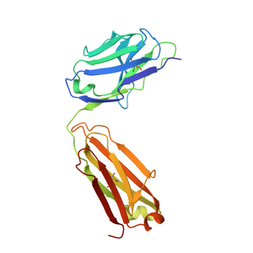
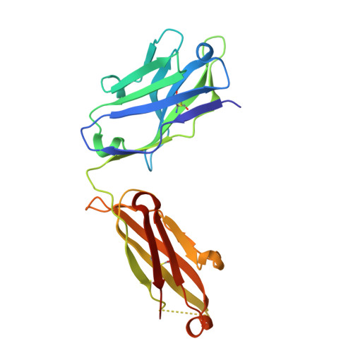
Human Rotavirus VP6-Specific Antibodies Mediate Intracellular Neutralization by Binding to a Quaternary Structure in the Transcriptional Pore.
Aiyegbo, M.S., Sapparapu, G., Spiller, B.W., Eli, I.M., Williams, D.R., Kim, R., Lee, D.E., Liu, T., Li, S., Woods, V.L., Nannemann, D.P., Meiler, J., Stewart, P.L., Crowe, J.E.(2013) PLoS One 8: e61101-e61101
- PubMed: 23671563
- DOI: https://doi.org/10.1371/journal.pone.0061101
- Primary Citation of Related Structures:
4HFW - PubMed Abstract:
Several live attenuated rotavirus (RV) vaccines have been licensed, but the mechanisms of protective immunity are still poorly understood. The most frequent human B cell response is directed to the internal protein VP6 on the surface of double-layered particles, which is normally exposed only in the intracellular environment. Here, we show that the canonical VP6 antibodies secreted by humans bind to such particles and inhibit viral transcription. Polymeric IgA RV antibodies mediated an inhibitory effect against virus replication inside cells during IgA transcytosis. We defined the recognition site on VP6 as a quaternary epitope containing a high density of charged residues. RV human mAbs appear to bind to a negatively-charged patch on the surface of the Type I channel in the transcriptionally active particle, and they sterically block the channel. This unique mucosal mechanism of viral neutralization, which is not apparent from conventional immunoassays, may contribute significantly to human immunity to RV.
Organizational Affiliation:
Department of Pathology, Microbiology and Immunology, Vanderbilt Medical Center, Nashville, Tennessee, USA.