4HD8

Crystal structure of human Sirt3 in complex with Fluor-de-Lys peptide and piceatannol


Experimental Data Snapshot

  • Method: X-RAY DIFFRACTION
  • Resolution: 2.30 Å
  • R-Value Free: 0.240 
  • R-Value Work: 0.174 
  • R-Value Observed: 0.177 

wwPDB Validation   3D Report Full Report


Ligand Structure Quality Assessment 


This is version 1.2 of the entry. See complete history


Literature

A molecular mechanism for direct sirtuin activation by resveratrol.

Gertz, M.Nguyen, G.T.Fischer, F.Suenkel, B.Schlicker, C.Franzel, B.Tomaschewski, J.Aladini, F.Becker, C.Wolters, D.Steegborn, C.

(2012) PLoS One 7: e49761-e49761

  • DOI: https://doi.org/10.1371/journal.pone.0049761
  • Primary Citation of Related Structures:  
    4HD8, 4HDA

  • PubMed Abstract: 

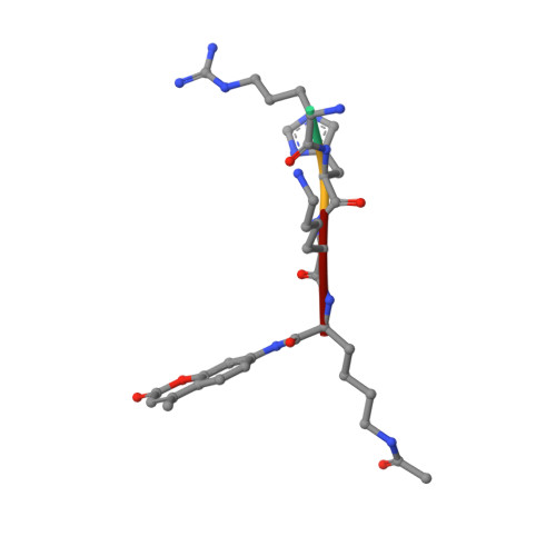
    Sirtuins are protein deacetylases regulating metabolism, stress responses, and aging processes, and they were suggested to mediate the lifespan extending effect of a low calorie diet. Sirtuin activation by the polyphenol resveratrol can mimic such lifespan extending effects and alleviate metabolic diseases. The mechanism of Sirtuin stimulation is unknown, hindering the development of improved activators. Here we show that resveratrol inhibits human Sirt3 and stimulates Sirt5, in addition to Sirt1, against fluorophore-labeled peptide substrates but also against peptides and proteins lacking the non-physiological fluorophore modification. We further present crystal structures of Sirt3 and Sirt5 in complex with fluorogenic substrate peptide and modulator. The compound acts as a top cover, closing the Sirtuin's polypeptide binding pocket and influencing details of peptide binding by directly interacting with this substrate. Our results provide a mechanism for the direct activation of Sirtuins by small molecules and suggest that activators have to be tailored to a specific Sirtuin/substrate pair.


  • Organizational Affiliation

    Department of Biochemistry, University of Bayreuth, Bayreuth, Germany.


Macromolecules
Find similar proteins by:  (by identity cutoff)  |  3D Structure
Entity ID: 1
MoleculeChains Sequence LengthOrganismDetailsImage
NAD-dependent protein deacetylase sirtuin-3, mitochondrial284Homo sapiensMutation(s): 0 
Gene Names: SIR2L3SIRT3
EC: 3.5.1
UniProt & NIH Common Fund Data Resources
Find proteins for Q9NTG7 (Homo sapiens)
Explore Q9NTG7 
Go to UniProtKB:  Q9NTG7
PHAROS:  Q9NTG7
GTEx:  ENSG00000142082 
Entity Groups  
Sequence Clusters30% Identity50% Identity70% Identity90% Identity95% Identity100% Identity
UniProt GroupQ9NTG7
Sequence Annotations
Expand
  • Reference Sequence

Find similar proteins by:  Sequence   |   3D Structure  

Entity ID: 2
MoleculeChains Sequence LengthOrganismDetailsImage
Fluor-de-Lys peptideB [auth F]4N/AMutation(s): 0 
Sequence Annotations
Expand
  • Reference Sequence
Small Molecules
Modified Residues  1 Unique
IDChains TypeFormula2D DiagramParent
FDL
Query on FDL
B [auth F]L-PEPTIDE LINKINGC18 H23 N3 O4LYS
Experimental Data & Validation

Experimental Data

  • Method: X-RAY DIFFRACTION
  • Resolution: 2.30 Å
  • R-Value Free: 0.240 
  • R-Value Work: 0.174 
  • R-Value Observed: 0.177 
  • Space Group: H 3 2
Unit Cell:
Length ( Å )Angle ( ˚ )
a = 114.6α = 90
b = 114.6β = 90
c = 123.7γ = 120
Software Package:
Software NamePurpose
XDSdata scaling
MOLREPphasing
REFMACrefinement
XDSdata reduction
XSCALEdata scaling

Structure Validation

View Full Validation Report


Ligand Structure Quality Assessment 


Entry History 

Deposition Data

Revision History  (Full details and data files)

  • Version 1.0: 2012-12-05
    Type: Initial release
  • Version 1.1: 2013-02-20
    Changes: Database references
  • Version 1.2: 2024-02-28
    Changes: Data collection, Database references, Derived calculations