


Structure of Staphylococcus aureus biotin protein ligase in complex with biotin acetylene
Yap, M., Wilce, M., Polyak, S, Soares da Costa, T., Tieu, W.To be published.
Experimental Data Snapshot
Entity ID: 1 | |||||
|---|---|---|---|---|---|
| Molecule | Chains | Sequence Length | Organism | Details | Image |
| Biotin-[acetyl-CoA-carboxylase] ligase | 328 | Staphylococcus aureus subsp. aureus ECT-R 2 | Mutation(s): 0 Gene Names: ECTR2_1310 EC: 6.3.4.15 | 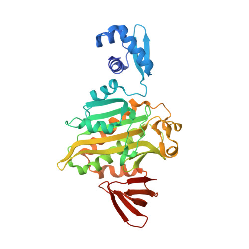 | |
Entity Groups | |||||
| Sequence Clusters | 30% Identity50% Identity70% Identity90% Identity95% Identity100% Identity | ||||
Sequence AnnotationsExpand | |||||
| |||||
| Ligands 1 Unique | |||||
|---|---|---|---|---|---|
| ID | Chains | Name / Formula / InChI Key | 2D Diagram | 3D Interactions | |
| BC4 Query on BC4 | B [auth A] | (3aS,4S,6aR)-4-(hex-5-yn-1-yl)tetrahydro-1H-thieno[3,4-d]imidazol-2(3H)-one C11 H16 N2 O S MGFIEEWTLUFGHD-GUBZILKMSA-N |  | ||
| Length ( Å ) | Angle ( ˚ ) |
|---|---|
| a = 93.02 | α = 90 |
| b = 93.02 | β = 90 |
| c = 129.94 | γ = 90 |
| Software Name | Purpose |
|---|---|
| XDS | data scaling |
| REFMAC | refinement |
RCSB PDB (citation) is hosted by
RCSB PDB is a member of the

