4H7A

Crystal structure of CasB from Thermus thermophilus


Experimental Data Snapshot

  • Method: X-RAY DIFFRACTION
  • Resolution: 2.60 Å
  • R-Value Free: 0.230 
  • R-Value Work: 0.173 
  • R-Value Observed: 0.176 

wwPDB Validation   3D Report Full Report


This is version 1.3 of the entry. See complete history


Literature

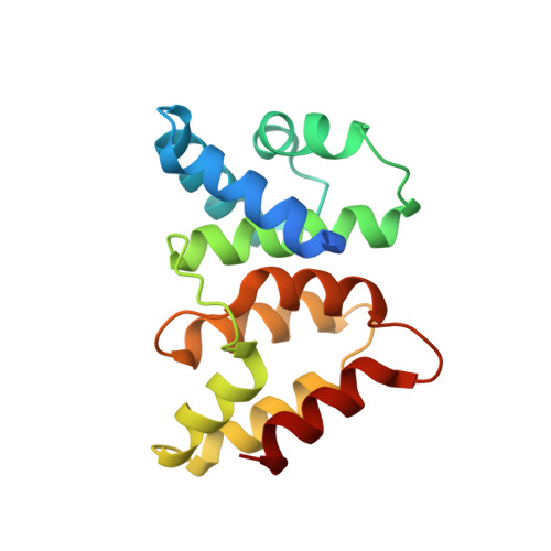
Nucleic acid binding surface and dimer interface revealed by CRISPR-associated CasB protein structures.

Nam, K.H.Huang, Q.Ke, A.

(2012) FEBS Lett 586: 3956-3961

  • DOI: https://doi.org/10.1016/j.febslet.2012.09.041
  • Primary Citation of Related Structures:  
    4H79, 4H7A

  • PubMed Abstract: 

    The CRISPR system is an adaptive RNA-based microbial immune system against invasive genetic elements. CasB is an essential protein component in Type I-E Cascade. Here, we characterize CasB proteins from three different organisms as non-specific nucleic acid binding proteins. The Thermobifida fusca CasB crystal structure reveals conserved positive surface charges, which we show are important for its nucleic acid binding function. EM docking reveals that CasB dimerization aligns individual nucleic acid binding surfaces into a curved, elongated binding surface inside Type I-E Cascade, consistent with the putative functions of CasB in ds-DNA recruitment and crRNA-DNA duplex formation steps.


  • Organizational Affiliation

    Department of Molecular Biology and Genetics, Cornell University, Ithaca, NY 14850, USA.


Macromolecules
Find similar proteins by:  (by identity cutoff)  |  3D Structure
Entity ID: 1
MoleculeChains Sequence LengthOrganismDetailsImage
CRISPR-associated protein Cse2
A, B
168Thermus thermophilus HB8Mutation(s): 0 
Gene Names: cse2TTHB189
UniProt
Find proteins for Q53VY0 (Thermus thermophilus (strain ATCC 27634 / DSM 579 / HB8))
Explore Q53VY0 
Go to UniProtKB:  Q53VY0
Entity Groups  
Sequence Clusters30% Identity50% Identity70% Identity90% Identity95% Identity100% Identity
UniProt GroupQ53VY0
Sequence Annotations
Expand
  • Reference Sequence
Experimental Data & Validation

Experimental Data

  • Method: X-RAY DIFFRACTION
  • Resolution: 2.60 Å
  • R-Value Free: 0.230 
  • R-Value Work: 0.173 
  • R-Value Observed: 0.176 
  • Space Group: P 43 21 2
Unit Cell:
Length ( Å )Angle ( ˚ )
a = 100.777α = 90
b = 100.777β = 90
c = 84.862γ = 90
Software Package:
Software NamePurpose
SCALEPACKdata scaling
MOLREPphasing
REFMACrefinement
PDB_EXTRACTdata extraction
HKL-2000data collection
HKL-2000data reduction
HKL-2000data scaling

Structure Validation

View Full Validation Report


Entry History 

Deposition Data

Revision History  (Full details and data files)

  • Version 1.0: 2012-10-03
    Type: Initial release
  • Version 1.1: 2012-11-28
    Changes: Database references
  • Version 1.2: 2017-10-25
    Changes: Author supporting evidence
  • Version 1.3: 2023-09-20
    Changes: Data collection, Database references, Refinement description