


Structure of anthranilate phosphoribosyl transferase from acinetobacter baylyi
Ponniah, K., Nigon, L.V., Anderson, B.F., Norris, G.E., Patrick, W.M.To be published.
Experimental Data Snapshot
wwPDB Validation 3D Report Full Report

Entity ID: 1 | |||||
|---|---|---|---|---|---|
| Molecule | Chains | Sequence Length | Organism | Details | Image |
| Anthranilate phosphoribosyltransferase | 353 | Acinetobacter baylyi ADP1 | Mutation(s): 0 Gene Names: trpD EC: 2.4.2.18 | 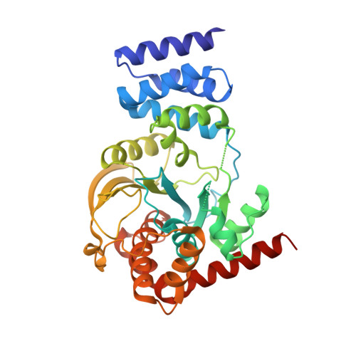 | |
UniProt | |||||
Find proteins for P00500 (Acinetobacter baylyi (strain ATCC 33305 / BD413 / ADP1)) Explore P00500 Go to UniProtKB: P00500 | |||||
Entity Groups | |||||
| Sequence Clusters | 30% Identity50% Identity70% Identity90% Identity95% Identity100% Identity | ||||
| UniProt Group | P00500 | ||||
Sequence AnnotationsExpand | |||||
| |||||
| Length ( Å ) | Angle ( ˚ ) |
|---|---|
| a = 59.58 | α = 90 |
| b = 70.1 | β = 90 |
| c = 171.41 | γ = 90 |
| Software Name | Purpose |
|---|---|
| CrystalClear | data collection |
| PHASER | phasing |
| PHENIX | refinement |
| xia2 | data reduction |
| XDS | data reduction |
| SCALA | data scaling |
RCSB PDB (citation) is hosted by
RCSB PDB is a member of the

