Mapping the Active Site Helix-to-Strand Conversion of CxxxxC Peroxiredoxin Q Enzymes.
Perkins, A., Gretes, M.C., Nelson, K.J., Poole, L.B., Karplus, P.A.(2012) Biochemistry 51: 7638-7650
- PubMed: 22928725
- DOI: https://doi.org/10.1021/bi301017s
- Primary Citation of Related Structures:
4G2E, 4GQC, 4GQF - PubMed Abstract:
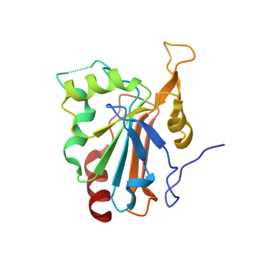
Peroxiredoxins (Prx) make up a family of enzymes that reduce peroxides using a peroxidatic cysteine residue; among these, members of the PrxQ subfamily are proposed to be the most ancestral-like yet are among the least characterized. In many PrxQ enzymes, a second "resolving" cysteine is located five residues downstream from the peroxidatic Cys, and these residues form a disulfide during the catalytic cycle. Here, we describe three hyperthermophilic PrxQ crystal structures originally determined by the RIKEN structural genomics group. We reprocessed the diffraction data and conducted further refinement to yield models with R(free) values lowered by 2.3-7.2% and resolution extended by 0.2-0.3 Å, making one, at 1.4 Å, one of the best resolved peroxiredoxins to date. Comparisons of two matched thiol and disulfide forms reveal that the active site conformational change required for disulfide formation involves a transition of ~20 residues from a pair of α-helices to a β-hairpin and 3(10)-helix. Each conformation has ~10 residues with a high level of disorder providing slack that allows the dramatic shift, and the two conformations are anchored to the protein core by distinct nonpolar side chains that fill three hydrophobic pockets. Sequence conservation patterns confirm the importance of these and a few additional residues for function. From a broader perspective, this study raises the provocative question of how to make use of the valuable information in the Protein Data Bank generated by structural genomics projects but not described in the literature, perhaps remaining unrecognized and certainly underutilized.
Organizational Affiliation:
Department of Biochemistry and Biophysics, Oregon State University, Corvallis, OR 97331, USA.