4G1O

Crystal structure of Newcastle disease virus matrix protein


Experimental Data Snapshot

  • Method: X-RAY DIFFRACTION
  • Resolution: 2.20 Å
  • R-Value Free: 0.280 
  • R-Value Work: 0.180 
  • R-Value Observed: 0.190 

wwPDB Validation   3D Report Full Report


This is version 1.2 of the entry. See complete history


Literature

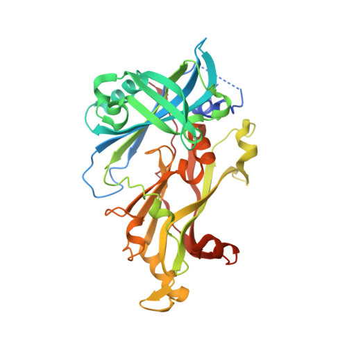
Structure and assembly of a paramyxovirus matrix protein.

Battisti, A.J.Meng, G.Winkler, D.C.McGinnes, L.W.Plevka, P.Steven, A.C.Morrison, T.G.Rossmann, M.G.

(2012) Proc Natl Acad Sci U S A 109: 13996-14000

  • DOI: https://doi.org/10.1073/pnas.1210275109
  • Primary Citation of Related Structures:  
    4G1G, 4G1L, 4G1O

  • PubMed Abstract: 

    Many pleomorphic, lipid-enveloped viruses encode matrix proteins that direct their assembly and budding, but the mechanism of this process is unclear. We have combined X-ray crystallography and cryoelectron tomography to show that the matrix protein of Newcastle disease virus, a paramyxovirus and relative of measles virus, forms dimers that assemble into pseudotetrameric arrays that generate the membrane curvature necessary for virus budding. We show that the glycoproteins are anchored in the gaps between the matrix proteins and that the helical nucleocapsids are associated in register with the matrix arrays. About 90% of virions lack matrix arrays, suggesting that, in agreement with previous biological observations, the matrix protein needs to dissociate from the viral membrane during maturation, as is required for fusion and release of the nucleocapsid into the host's cytoplasm. Structure and sequence conservation imply that other paramyxovirus matrix proteins function similarly.


  • Organizational Affiliation

    Department of Biological Sciences, Purdue University, West Lafayette, IN 47907-2032, USA.


Macromolecules
Find similar proteins by:  (by identity cutoff)  |  3D Structure
Entity ID: 1
MoleculeChains Sequence LengthOrganismDetailsImage
Matrix protein
A, B
364Newcastle disease virus (STRAIN AUSTRALIA-VICTORIA/32)Mutation(s): 0 
Gene Names: M
UniProt
Find proteins for P11206 (Newcastle disease virus (strain Chicken/Australia-Victoria/32))
Explore P11206 
Go to UniProtKB:  P11206
Entity Groups  
Sequence Clusters30% Identity50% Identity70% Identity90% Identity95% Identity100% Identity
UniProt GroupP11206
Sequence Annotations
Expand
  • Reference Sequence
Experimental Data & Validation

Experimental Data

  • Method: X-RAY DIFFRACTION
  • Resolution: 2.20 Å
  • R-Value Free: 0.280 
  • R-Value Work: 0.180 
  • R-Value Observed: 0.190 
  • Space Group: C 1 2 1
Unit Cell:
Length ( Å )Angle ( ˚ )
a = 163.311α = 90
b = 46.987β = 132.05
c = 117.41γ = 90
Software Package:
Software NamePurpose
MAR345dtbdata collection
MOLREPphasing
REFMACrefinement
HKL-2000data reduction
HKL-2000data scaling

Structure Validation

View Full Validation Report


Entry History 

Deposition Data

Revision History  (Full details and data files)

  • Version 1.0: 2012-08-22
    Type: Initial release
  • Version 1.1: 2012-09-12
    Changes: Database references
  • Version 1.2: 2024-02-28
    Changes: Data collection, Database references