4FT8

E. coli Catabolite Activator Protein with Cobalt and Sulfate Ligands


Experimental Data Snapshot

  • Method: X-RAY DIFFRACTION
  • Resolution: 1.97 Å
  • R-Value Free: 0.230 
  • R-Value Work: 0.196 
  • R-Value Observed: 0.198 

wwPDB Validation   3D Report Full Report


Ligand Structure Quality Assessment 


This is version 1.3 of the entry. See complete history


Literature

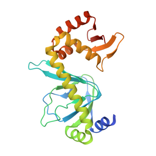
Structure of catabolite activator protein with cobalt(II) and sulfate.

Rao, R.R.Lawson, C.L.

(2014) Acta Crystallogr Sect F Struct Biol Cryst Commun 70: 560-563

  • DOI: https://doi.org/10.1107/S2053230X14005366
  • Primary Citation of Related Structures:  
    4FT8

  • PubMed Abstract: 

    The crystal structure of cyclic AMP-catabolite activator protein (CAP) from Escherichia coli containing cobalt(II) chloride and ammonium sulfate is reported at 1.97 Å resolution. Each of the two CAP subunits in the asymmetric unit binds one cobalt(II) ion, in each case coordinated by N-terminal domain residues His19, His21 and Glu96 plus an additional acidic residue contributed via a crystal contact. The three identified N-terminal domain cobalt-binding residues are part of a region of CAP that is important for transcription activation at class II CAP-dependent promoters. Sulfate anions mediate additional crystal lattice contacts and occupy sites corresponding to DNA backbone phosphate positions in CAP-DNA complex structures.


  • Organizational Affiliation

    Department of Chemistry and Chemical Biology, Rutgers University, Piscataway, NJ 08854, USA.


Macromolecules
Find similar proteins by:  (by identity cutoff)  |  3D Structure
Entity ID: 1
MoleculeChains Sequence LengthOrganismDetailsImage
Catabolite gene activator
A, B
209Escherichia coli K-12Mutation(s): 0 
Gene Names: b3357capcrpcsmJW5702
UniProt
Find proteins for P0ACJ8 (Escherichia coli (strain K12))
Explore P0ACJ8 
Go to UniProtKB:  P0ACJ8
Entity Groups  
Sequence Clusters30% Identity50% Identity70% Identity90% Identity95% Identity100% Identity
UniProt GroupP0ACJ8
Sequence Annotations
Expand
  • Reference Sequence
Small Molecules
Ligands 3 Unique
IDChains Name / Formula / InChI Key2D Diagram3D Interactions
CMP
Query on CMP

Download Ideal Coordinates CCD File 
C [auth A],
K [auth B]
ADENOSINE-3',5'-CYCLIC-MONOPHOSPHATE
C10 H12 N5 O6 P
IVOMOUWHDPKRLL-KQYNXXCUSA-N
SO4
Query on SO4

Download Ideal Coordinates CCD File 
D [auth A]
E [auth A]
F [auth A]
G [auth A]
H [auth A]
D [auth A],
E [auth A],
F [auth A],
G [auth A],
H [auth A],
I [auth A],
L [auth B],
M [auth B],
N [auth B],
O [auth B],
P [auth B],
Q [auth B],
R [auth B]
SULFATE ION
O4 S
QAOWNCQODCNURD-UHFFFAOYSA-L
CO
Query on CO

Download Ideal Coordinates CCD File 
J [auth A],
S [auth B]
COBALT (II) ION
Co
XLJKHNWPARRRJB-UHFFFAOYSA-N
Experimental Data & Validation

Experimental Data

  • Method: X-RAY DIFFRACTION
  • Resolution: 1.97 Å
  • R-Value Free: 0.230 
  • R-Value Work: 0.196 
  • R-Value Observed: 0.198 
  • Space Group: P 21 21 2
Unit Cell:
Length ( Å )Angle ( ˚ )
a = 104.143α = 90
b = 108.452β = 90
c = 44.32γ = 90
Software Package:
Software NamePurpose
DENZOdata reduction
SCALEPACKdata scaling
PHENIXrefinement
PDB_EXTRACTdata extraction
CBASSdata collection
PHASERphasing

Structure Validation

View Full Validation Report


Ligand Structure Quality Assessment 


Entry History 

Deposition Data

Revision History  (Full details and data files)

  • Version 1.0: 2013-12-18
    Type: Initial release
  • Version 1.1: 2014-05-07
    Changes: Database references
  • Version 1.2: 2014-09-24
    Changes: Database references
  • Version 1.3: 2023-09-13
    Changes: Data collection, Database references, Derived calculations, Refinement description