Evidence for a Dual Role of an Active Site Histidine in alpha-Amino-beta-Carboxymuconate-epsilon-Semialdehyde Decarboxylase
Huo, L., Fielding, A.J., Chen, Y., Li, T., Iwaki, H., Hosler, J.P., Chen, L., Hasegawa, Y., Que, L., Liu, A.(2012) Biochemistry 51: 5811-5821
- PubMed: 22746257
- DOI: https://doi.org/10.1021/bi300635b
- Primary Citation of Related Structures:
4EPK, 4ERA, 4ERG, 4ERI - PubMed Abstract:
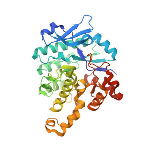
The previously reported crystal structures of α-amino-β-carboxymuconate-ε-semialdehyde decarboxylase (ACMSD) show a five-coordinate Zn(II)(His)(3)(Asp)(OH(2)) active site. The water ligand is H-bonded to a conserved His228 residue adjacent to the metal center in ACMSD from Pseudomonas fluorescens (PfACMSD). Site-directed mutagenesis of His228 to tyrosine and glycine in this study results in a complete or significant loss of activity. Metal analysis shows that H228Y and H228G contain iron rather than zinc, indicating that this residue plays a role in the metal selectivity of the protein. As-isolated H228Y displays a blue color, which is not seen in wild-type ACMSD. Quinone staining and resonance Raman analyses indicate that the blue color originates from Fe(III)-tyrosinate ligand-to-metal charge transfer. Co(II)-substituted H228Y ACMSD is brown in color and exhibits an electron paramagnetic resonance spectrum showing a high-spin Co(II) center with a well-resolved (59)Co (I = 7/2) eight-line hyperfine splitting pattern. The X-ray crystal structures of as-isolated Fe-H228Y (2.8 Å) and Co-substituted (2.4 Å) and Zn-substituted H228Y (2.0 Å resolution) support the spectroscopic assignment of metal ligation of the Tyr228 residue. The crystal structure of Zn-H228G (2.6 Å) was also determined. These four structures show that the water ligand present in WT Zn-ACMSD is either missing (Fe-H228Y, Co-H228Y, and Zn-H228G) or disrupted (Zn-H228Y) in response to the His228 mutation. Together, these results highlight the importance of His228 for PfACMSD's metal specificity as well as maintaining a water molecule as a ligand of the metal center. His228 is thus proposed to play a role in activating the metal-bound water ligand for subsequent nucleophilic attack on the substrate.
Organizational Affiliation:
Department of Chemistry and Center for Diagnostics and Therapeutics, Georgia State University, P.O. Box 4098, Atlanta, GA 30303, USA.