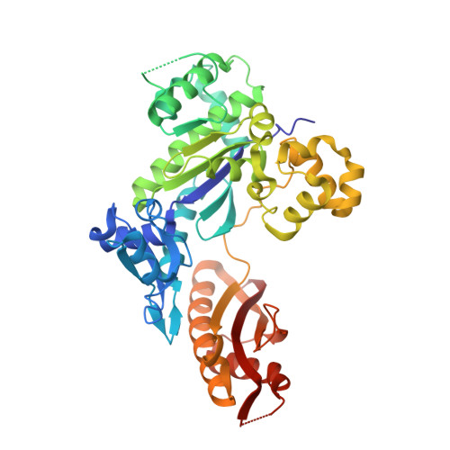
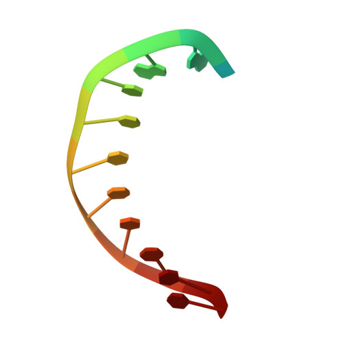
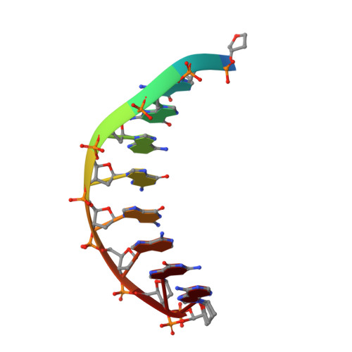
4EEY

Crystal structure of human DNA polymerase eta in ternary complex with a cisplatin DNA adduct


Experimental Data Snapshot

  • Method: X-RAY DIFFRACTION
  • Resolution: 2.32 Å
  • R-Value Free: 0.233 
  • R-Value Work: 0.178 
  • R-Value Observed: 0.182 

wwPDB Validation   3D Report Full Report


Ligand Structure Quality Assessment 


This is version 1.2 of the entry. See complete history


Literature

Structural basis for cisplatin DNA damage tolerance by human polymerase {eta} during cancer chemotherapy.

Ummat, A.Rechkoblit, O.Jain, R.Roy Choudhury, J.Johnson, R.E.Silverstein, T.D.Buku, A.Lone, S.Prakash, L.Prakash, S.Aggarwal, A.K.

(2012) Nat Struct Mol Biol 19: 628-632

  • DOI: https://doi.org/10.1038/nsmb.2295
  • Primary Citation of Related Structures:  
    4EEY

  • PubMed Abstract: 

    A major clinical problem in the use of cisplatin to treat cancers is tumor resistance. DNA polymerase η (Pol-η) is a crucial polymerase that allows cancer cells to cope with the cisplatin-DNA adducts that are formed during chemotherapy. We present here a structure of human Pol-η inserting deoxycytidine triphosphate (dCTP) opposite a cisplatin intrastrand cross-link (PtGpG). We show that the specificity of human Pol-η for PtGpG derives from an active site that is open to permit Watson-Crick geometry of the nascent PtGpG-dCTP base pair and to accommodate the lesion without steric hindrance. This specificity is augmented by the residues Gln38 and Ser62, which interact with PtGpG, and Arg61, which interacts with the incoming dCTP. Collectively, the structure provides a basis for understanding how Pol-η in human cells can tolerate the DNA damage caused by cisplatin chemotherapy and offers a framework for the design of inhibitors in cancer therapy.


  • Organizational Affiliation

    Department of Structural and Chemical Biology, Mount Sinai School of Medicine, New York, New York, USA.


Macromolecules

Find similar proteins by:  (by identity cutoff)  |  3D Structure
Entity ID: 1
MoleculeChains Sequence LengthOrganismDetailsImage
DNA polymerase eta435Homo sapiensMutation(s): 1 
Gene Names: POLHRAD30RAD30AXPV
EC: 2.7.7.7
UniProt & NIH Common Fund Data Resources
Find proteins for Q9Y253 (Homo sapiens)
Explore Q9Y253 
Go to UniProtKB:  Q9Y253
PHAROS:  Q9Y253
GTEx:  ENSG00000170734 
Entity Groups  
Sequence Clusters30% Identity50% Identity70% Identity90% Identity95% Identity100% Identity
UniProt GroupQ9Y253
Sequence Annotations
Expand
  • Reference Sequence

Find similar nucleic acids by:  Sequence   |   3D Structure  

Entity ID: 2
MoleculeChains LengthOrganismImage
5'-D(*CP*TP*TP*GP*GP*TP*CP*TP*CP*CP*TP*CP*C)-3'B [auth T]13N/A
Sequence Annotations
Expand
  • Reference Sequence

Find similar nucleic acids by:  Sequence   |   3D Structure  

Entity ID: 3
MoleculeChains LengthOrganismImage
5'-D(*TP*GP*GP*AP*GP*GP*AP*GP*A)-3'C [auth P]9N/A
Sequence Annotations
Expand
  • Reference Sequence
Experimental Data & Validation

Experimental Data

  • Method: X-RAY DIFFRACTION
  • Resolution: 2.32 Å
  • R-Value Free: 0.233 
  • R-Value Work: 0.178 
  • R-Value Observed: 0.182 
  • Space Group: P 61
Unit Cell:
Length ( Å )Angle ( ˚ )
a = 98.5α = 90
b = 98.5β = 90
c = 82.603γ = 120
Software Package:
Software NamePurpose
CBASSdata collection
PHASERphasing
REFMACrefinement
HKL-2000data reduction
HKL-2000data scaling

Structure Validation

View Full Validation Report


Ligand Structure Quality Assessment 


Entry History 

Deposition Data

Revision History  (Full details and data files)

  • Version 1.0: 2012-05-09
    Type: Initial release
  • Version 1.1: 2013-06-19
    Changes: Database references
  • Version 1.2: 2023-09-13
    Changes: Data collection, Database references, Derived calculations, Refinement description