4DLD

Crystal structure of the GluK1 ligand-binding domain (S1S2) in complex with the antagonist (S)-2-amino-3-(2-(2-carboxyethyl)-5-chloro-4-nitrophenyl)propionic acid at 2.0 A resolution


Experimental Data Snapshot

  • Method: X-RAY DIFFRACTION
  • Resolution: 2.00 Å
  • R-Value Free: 0.246 
  • R-Value Work: 0.186 
  • R-Value Observed: 0.189 

wwPDB Validation   3D Report Full Report


Ligand Structure Quality Assessment 


This is version 1.2 of the entry. See complete history


Literature

Structural and pharmacological characterization of phenylalanine-based AMPA receptor antagonists at kainate receptors

Venskutonyte, R.Frydenvang, K.Valades, E.A.Szymanska, E.Johansen, T.N.Kastrup, J.S.Pickering, D.S.

(2012) ChemMedChem 7: 1793-1798

  • DOI: https://doi.org/10.1002/cmdc.201100599
  • Primary Citation of Related Structures:  
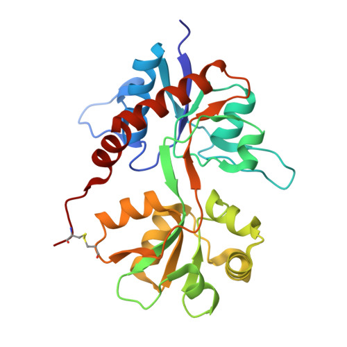
    4DLD

  • PubMed Abstract: 

    Continued efforts into the discovery of ligands that target ionotropic glutamate receptors (iGluRs) are important for studies of the physiological roles of the various iGluR subtypes as well as for the search for drugs that can be used in the treatment of diseases of the central nervous system. A new series of phenylalanine derivatives that target iGluRs was reported to bind AMPA receptors. Herein we report our studies of these compounds at the kainate receptors GluK1-3. Several compounds bind with micromolar affinity at GluK1 and GluK3, but do not bind GluK2. The crystal structure of the most potent compound in the ligand binding domain of GluK1 revealed different modes of binding to GluK1 and GluA2, due primarily to residues Ser741 (GluK1) and Met729 (GluA2). The compound was shown to be slightly more potent at GluK1 than at AMPA receptors and to induce a domain closure similar to that observed in GluK1 structures with partial agonists.


  • Organizational Affiliation

    Department Drug Design and Pharmacology, Faculty of Health and Medical Sciences, University of Copenhagen, Universitetsparken 2, 2100 Copenhagen, Denmark.


Macromolecules
Find similar proteins by:  (by identity cutoff)  |  3D Structure
Entity ID: 1
MoleculeChains Sequence LengthOrganismDetailsImage
Glutamate receptor, ionotropic kainate 1
A, B
257Rattus norvegicusMutation(s): 0 
Gene Names: Grik1
UniProt
Find proteins for P22756 (Rattus norvegicus)
Explore P22756 
Go to UniProtKB:  P22756
Entity Groups  
Sequence Clusters30% Identity50% Identity70% Identity90% Identity95% Identity100% Identity
UniProt GroupP22756
Sequence Annotations
Expand
  • Reference Sequence
Small Molecules
Binding Affinity Annotations 
IDSourceBinding Affinity
TZG PDBBind:  4DLD Ki: 1500 (nM) from 1 assay(s)
BindingDB:  4DLD Ki: 3000 (nM) from 1 assay(s)
Binding MOAD:  4DLD Ki: 1500 (nM) from 1 assay(s)
Experimental Data & Validation

Experimental Data

  • Method: X-RAY DIFFRACTION
  • Resolution: 2.00 Å
  • R-Value Free: 0.246 
  • R-Value Work: 0.186 
  • R-Value Observed: 0.189 
  • Space Group: P 41 21 2
Unit Cell:
Length ( Å )Angle ( ˚ )
a = 69.32α = 90
b = 69.32β = 90
c = 234.97γ = 90
Software Package:
Software NamePurpose
MOSFLMdata reduction
SCALAdata scaling
PHASERphasing
PHENIXrefinement
PDB_EXTRACTdata extraction

Structure Validation

View Full Validation Report


Ligand Structure Quality Assessment 


Entry History 

Deposition Data

Revision History  (Full details and data files)

  • Version 1.0: 2012-10-10
    Type: Initial release
  • Version 1.1: 2017-08-16
    Changes: Data collection, Refinement description, Source and taxonomy
  • Version 1.2: 2023-11-08
    Changes: Data collection, Database references, Derived calculations, Refinement description