Dimerization of Matrix Protein is Required for Budding of Respiratory Syncytial Virus.
Forster, A., Maertens, G.N., Farrell, P.J., Bajorek, M.(2015) J Virol 89: 4624
- PubMed: 25673702
- DOI: https://doi.org/10.1128/JVI.03500-14
- Primary Citation of Related Structures:
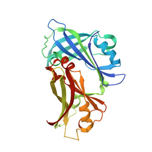
4D4T, 4V23 - PubMed Abstract:
Respiratory syncytial virus (RSV) infects epithelial cells of the respiratory tract and is a major cause of bronchiolitis and pneumonia in children and the elderly. The virus assembles and buds through the plasma membrane, forming elongated membrane filaments, but details of how this happens remain obscure. Oligomerization of the matrix protein (M) is a key step in the process of assembly and infectious virus production. In addition, it was suggested to affect the conformation of the fusion protein, the major current target for RSV antivirals, in the mature virus. The structure and assembly of M are thus key parameters in the RSV antiviral development strategy. The structure of RSV M was previously published as a monomer. Other paramyxovirus M proteins have been shown to dimerize, and biochemical data suggest that RSV M also dimerizes. Here, using size exclusion chromatography-multiangle laser light scattering, we show that the protein is dimeric in solution. We also crystallized M in two crystal forms and show that it assembles into equivalent dimers in both lattices. Dimerization interface mutations destabilize the M dimer in vitro. To assess the biological relevance of dimerization, we used confocal imaging to show that dimerization interface mutants of M fail to assemble into viral filaments on the plasma membrane. Additionally, budding and release of virus-like particles are prevented in M mutants that fail to form filaments. Importantly, we show that M is biologically active as a dimer and that the switch from M dimers to higher-order oligomers triggers viral filament assembly and virus production.
Organizational Affiliation:
Centre for Structural Biology, Department of Life Sciences, Imperial College London, London, United Kingdom.