4CZH

C. crescentus MreB, single filament, ADP, MP265 inhibitor


Experimental Data Snapshot

  • Method: X-RAY DIFFRACTION
  • Resolution: 1.64 Å
  • R-Value Free: 0.232 
  • R-Value Work: 0.201 
  • R-Value Observed: 0.202 

wwPDB Validation   3D Report Full Report


Ligand Structure Quality Assessment 


This is version 1.2 of the entry. See complete history


Literature

Bacterial Actin Mreb Forms Antiparallel Double Filaments.

Van Den Ent, F.Izore, T.Bharat, T.A.Johnson, C.M.Lowe, J.

(2014) Elife 3: 02634

  • DOI: https://doi.org/10.7554/eLife.02634
  • Primary Citation of Related Structures:  
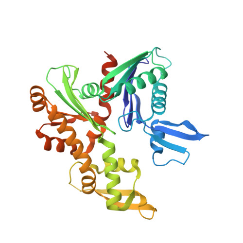
    4CZE, 4CZF, 4CZG, 4CZH, 4CZI, 4CZJ, 4CZK, 4CZL, 4CZM

  • PubMed Abstract: 

    Filaments of all actin-like proteins known to date are assembled from pairs of protofilaments that are arranged in a parallel fashion, generating polarity. In this study, we show that the prokaryotic actin homologue MreB forms pairs of protofilaments that adopt an antiparallel arrangement in vitro and in vivo. We provide an atomic view of antiparallel protofilaments of Caulobacter MreB as apparent from crystal structures. We show that a protofilament doublet is essential for MreB's function in cell shape maintenance and demonstrate by in vivo site-specific cross-linking the antiparallel orientation of MreB protofilaments in E. coli. 3D cryo-EM shows that pairs of protofilaments of Caulobacter MreB tightly bind to membranes. Crystal structures of different nucleotide and polymerisation states of Caulobacter MreB reveal conserved conformational changes accompanying antiparallel filament formation. Finally, the antimicrobial agents A22/MP265 are shown to bind close to the bound nucleotide of MreB, presumably preventing nucleotide hydrolysis and destabilising double protofilaments.DOI: http://dx.doi.org/10.7554/eLife.02634.001.


  • Organizational Affiliation

    Structural Studies Division, Medical Research Council - Laboratory of Molecular Biology, Cambridge, United Kingdom fent@mrc-lmb.cam.ac.uk.


Macromolecules
Find similar proteins by:  (by identity cutoff)  |  3D Structure
Entity ID: 1
MoleculeChains Sequence LengthOrganismDetailsImage
ROD SHAPE-DETERMINING PROTEIN MREB348Caulobacter vibrioidesMutation(s): 2 
UniProt
Find proteins for A0A0H3C7V4 (Caulobacter vibrioides (strain NA1000 / CB15N))
Explore A0A0H3C7V4 
Go to UniProtKB:  A0A0H3C7V4
Entity Groups  
Sequence Clusters30% Identity50% Identity70% Identity90% Identity95% Identity100% Identity
UniProt GroupA0A0H3C7V4
Sequence Annotations
Expand
  • Reference Sequence
Small Molecules
Experimental Data & Validation

Experimental Data

  • Method: X-RAY DIFFRACTION
  • Resolution: 1.64 Å
  • R-Value Free: 0.232 
  • R-Value Work: 0.201 
  • R-Value Observed: 0.202 
  • Space Group: P 1 21 1
Unit Cell:
Length ( Å )Angle ( ˚ )
a = 50.618α = 90
b = 73.739β = 102.38
c = 53.7γ = 90
Software Package:
Software NamePurpose
PHENIXrefinement
XDSdata reduction
SCALAdata scaling
PHASERphasing

Structure Validation

View Full Validation Report


Ligand Structure Quality Assessment 


Entry History 

Deposition Data

Revision History  (Full details and data files)

  • Version 1.0: 2014-06-11
    Type: Initial release
  • Version 1.1: 2014-06-25
    Changes: Atomic model, Database references, Derived calculations, Other
  • Version 1.2: 2023-12-20
    Changes: Data collection, Database references, Derived calculations, Other, Refinement description