4CO1

Structure of PII signaling protein GlnZ from Azospirillum brasilense in complex with adenosine diphosphate


Experimental Data Snapshot

  • Method: X-RAY DIFFRACTION
  • Resolution: 1.50 Å
  • R-Value Free: 0.171 
  • R-Value Work: 0.139 
  • R-Value Observed: 0.146 

wwPDB Validation   3D Report Full Report


Ligand Structure Quality Assessment 


This is version 1.5 of the entry. See complete history


Literature

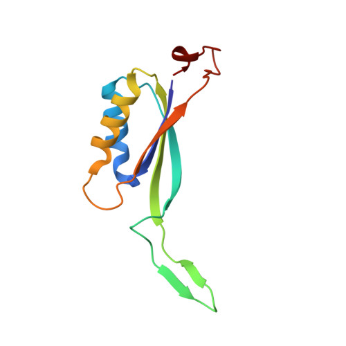
Structure and Thermodynamics of Effector Molecule Binding to the Nitrogen Signal Transduction Pii Protein Glnz from Azospirillum Brasilense.

Truan, D.Bjelic, S.Li, X.Winkler, F.K.

(2014) J Mol Biol 426: 2783

  • DOI: https://doi.org/10.1016/j.jmb.2014.05.008
  • Primary Citation of Related Structures:  
    4CNY, 4CNZ, 4CO0, 4CO1, 4CO2, 4CO3, 4CO4, 4CO5

  • PubMed Abstract: 

    The trimeric PII signal transduction proteins regulate the function of a variety of target proteins predominantly involved in nitrogen metabolism. ATP, ADP and 2-oxoglutarate (2-OG) are key effector molecules influencing PII binding to targets. Studies of PII proteins have established that the 20-residue T-loop plays a central role in effector sensing and target binding. However, the specific effects of effector binding on T-loop conformation have remained poorly documented. We present eight crystal structures of the Azospirillum brasilense PII protein GlnZ, six of which are cocrystallized and liganded with ADP or ATP. We find that interaction with the diphosphate moiety of bound ADP constrains the N-terminal part of the T-loop in a characteristic way that is maintained in ADP-promoted complexes with target proteins. In contrast, the interactions with the triphosphate moiety in ATP complexes are much more variable and no single predominant interaction mode is apparent except for the ternary MgATP/2-OG complex. These conclusions can be extended to most investigated PII proteins of the GlnB/GlnK subfamily. Unlike reported for other PII proteins, microcalorimetry reveals no cooperativity between the three binding sites of GlnZ trimers for any of the three effectors under carefully controlled experimental conditions.


  • Organizational Affiliation

    Macromolecular Crystallography, Swiss Light Source, CH-5232 Villigen PSI, Switzerland.


Macromolecules
Find similar proteins by:  (by identity cutoff)  |  3D Structure
Entity ID: 1
MoleculeChains Sequence LengthOrganismDetailsImage
PII-LIKE PROTEIN PZ
A, B
112Azospirillum brasilenseMutation(s): 0 
UniProt
Find proteins for P70731 (Azospirillum brasilense)
Explore P70731 
Go to UniProtKB:  P70731
Entity Groups  
Sequence Clusters30% Identity50% Identity70% Identity90% Identity95% Identity100% Identity
UniProt GroupP70731
Sequence Annotations
Expand
  • Reference Sequence
Small Molecules
Binding Affinity Annotations 
IDSourceBinding Affinity
ADP Binding MOAD:  4CO1 Kd: 2.00e+4 (nM) from 1 assay(s)
Experimental Data & Validation

Experimental Data

  • Method: X-RAY DIFFRACTION
  • Resolution: 1.50 Å
  • R-Value Free: 0.171 
  • R-Value Work: 0.139 
  • R-Value Observed: 0.146 
  • Space Group: H 3
Unit Cell:
Length ( Å )Angle ( ˚ )
a = 89.43α = 90
b = 89.43β = 90
c = 67.37γ = 120
Software Package:
Software NamePurpose
PHENIXrefinement
XDSdata reduction
XSCALEdata scaling
PHASERphasing

Structure Validation

View Full Validation Report


Ligand Structure Quality Assessment 


Entry History 

Deposition Data

Revision History  (Full details and data files)

  • Version 1.0: 2014-05-28
    Type: Initial release
  • Version 1.1: 2014-06-04
    Changes: Database references
  • Version 1.2: 2014-07-30
    Changes: Database references
  • Version 1.3: 2019-01-30
    Changes: Data collection, Experimental preparation
  • Version 1.4: 2019-02-06
    Changes: Data collection, Experimental preparation
  • Version 1.5: 2023-12-20
    Changes: Data collection, Database references, Derived calculations, Other, Refinement description