Crystal Structure of the Human Short Coiled Coil Protein and Insights Into Scoc-Fez1 Complex Formation.
Behrens, C., Binotti, B., Schmidt, C., Robinson, C.V., Chua, J.J.E., Kuhnel, K.(2013) PLoS One 8: 76355
- PubMed: 24098481
- DOI: https://doi.org/10.1371/journal.pone.0076355
- Primary Citation of Related Structures:
4BWD - PubMed Abstract:
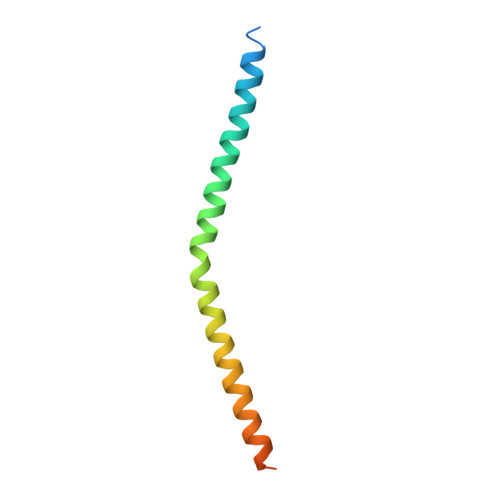
The short coiled coil protein (SCOC) forms a complex with fasciculation and elongation protein zeta 1 (FEZ1). This complex is involved in autophagy regulation. We determined the crystal structure of the coiled coil domain of human SCOC at 2.7 Å resolution. SCOC forms a parallel left handed coiled coil dimer. We observed two distinct dimers in the crystal structure, which shows that SCOC is conformationally flexible. This plasticity is due to the high incidence of polar and charged residues at the core a/d-heptad positions. We prepared two double mutants, where these core residues were mutated to either leucines or valines (E93V/K97L and N125L/N132V). These mutations led to a dramatic increase in stability and change of oligomerisation state. The oligomerisation state of the mutants was characterized by multi-angle laser light scattering and native mass spectrometry measurements. The E93V/K97 mutant forms a trimer and the N125L/N132V mutant is a tetramer. We further demonstrate that SCOC forms a stable homogeneous complex with the coiled coil domain of FEZ1. SCOC dimerization and the SCOC surface residue R117 are important for this interaction.
Organizational Affiliation:
Department of Neurobiology, Max-Planck-Institute for Biophysical Chemistry, Göttingen, Germany.