


4BTX
Crystal structure of human vascular adhesion protein-1 in complex with pyridazinone inhibitors
- PDB DOI: https://doi.org/10.2210/pdb4BTX/pdb
- Classification: OXIDOREDUCTASE
- Organism(s): Homo sapiens
- Mutation(s): No
- Deposited: 2013-06-19 Released: 2013-12-18
Experimental Data Snapshot
- Method: X-RAY DIFFRACTION
- Resolution: 2.78 Å
- R-Value Free: 0.237
- R-Value Work: 0.181
- R-Value Observed: 0.183

This is version 2.1 of the entry. See complete history.

Macromolecules
Find similar proteins by:
(by identity cutoff) | 3D Structure
Entity ID: 1 | |||||
|---|---|---|---|---|---|
| Molecule | Chains | Sequence Length | Organism | Details | Image |
| MEMBRANE PRIMARY AMINE OXIDASE | 737 | Homo sapiens | Mutation(s): 0 EC: 1.4.3.21 | 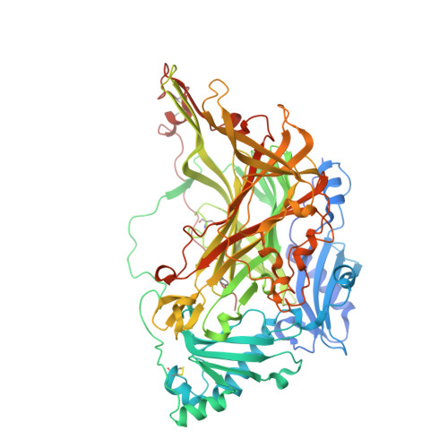 | |
UniProt & NIH Common Fund Data Resources | |||||
Find proteins for Q16853 (Homo sapiens) Explore Q16853 Go to UniProtKB: Q16853 | |||||
PHAROS: Q16853 GTEx: ENSG00000131471 | |||||
Entity Groups | |||||
| Sequence Clusters | 30% Identity50% Identity70% Identity90% Identity95% Identity100% Identity | ||||
| UniProt Group | Q16853 | ||||
Sequence AnnotationsExpand | |||||
| |||||
Oligosaccharides


Small Molecules
| Ligands 5 Unique | |||||
|---|---|---|---|---|---|
| ID | Chains | Name / Formula / InChI Key | 2D Diagram | 3D Interactions | |
| WF8 Query on WF8 | K [auth A], T [auth B] | 5-isopropylamino-2-phenyl-6-(1H-1,2,4-triazol-5-yl)-3(2H)-pyridazinone C15 H16 N6 O VZIFGHQBDGDZHH-UHFFFAOYSA-N |  | ||
| NAG Query on NAG | H [auth A] I [auth A] J [auth A] P [auth B] Q [auth B] | 2-acetamido-2-deoxy-beta-D-glucopyranose C8 H15 N O6 OVRNDRQMDRJTHS-FMDGEEDCSA-N |  | ||
| MAN Query on MAN | O [auth B] | alpha-D-mannopyranose C6 H12 O6 WQZGKKKJIJFFOK-PQMKYFCFSA-N |  | ||
| CU Query on CU | E [auth A], L [auth B] | COPPER (II) ION Cu JPVYNHNXODAKFH-UHFFFAOYSA-N |  | ||
| CA Query on CA | F [auth A], G [auth A], M [auth B], N [auth B] | CALCIUM ION Ca BHPQYMZQTOCNFJ-UHFFFAOYSA-N |  | ||
| Modified Residues 1 Unique | |||||
|---|---|---|---|---|---|
| ID | Chains | Type | Formula | 2D Diagram | Parent |
| TPQ Query on TPQ | A, B | L-PEPTIDE LINKING | C9 H9 N O5 |  | TYR |
Experimental Data & Validation
Experimental Data
- Method: X-RAY DIFFRACTION
- Resolution: 2.78 Å
- R-Value Free: 0.237
- R-Value Work: 0.181
- R-Value Observed: 0.183
- Space Group: P 65 2 2
Unit Cell:
| Length ( Å ) | Angle ( ˚ ) |
|---|---|
| a = 226.35 | α = 90 |
| b = 226.35 | β = 90 |
| c = 217.226 | γ = 120 |
| Software Name | Purpose |
|---|---|
| HKL | data reduction |
| HKL | data scaling |
| CCP4 | phasing |
| MOLREP | phasing |
| REFMAC | refinement |
Entry History
Deposition Data
- Released Date: 2013-12-18 Deposition Author(s): Bligt-Linden, E., Pihlavisto, M., Szatmari, I., Otwinowski, Z., Smith, D.J., Lazar, L., Fulop, F., Salminen, T.A.
Revision History (Full details and data files)
- Version 1.0: 2013-12-18
Type: Initial release - Version 1.1: 2014-01-15
Changes: Database references - Version 2.0: 2020-07-29
Type: Remediation
Reason: Carbohydrate remediation
Changes: Advisory, Atomic model, Data collection, Derived calculations, Other, Structure summary - Version 2.1: 2023-12-20
Changes: Data collection, Database references, Refinement description, Structure summary
