4BML

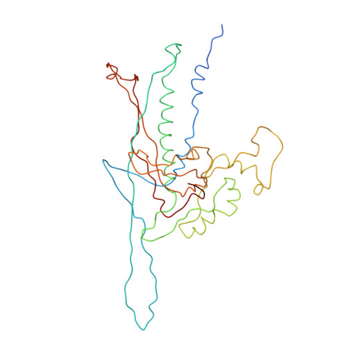
C-alpha backbone trace of major capsid protein gp39 found in marine virus Syn5.


Experimental Data Snapshot

  • Method: ELECTRON MICROSCOPY
  • Resolution: 4.70 Å
  • Aggregation State: PARTICLE 
  • Reconstruction Method: SINGLE PARTICLE 

wwPDB Validation   3D Report Full Report


This is version 1.5 of the entry. See complete history


Literature

Protruding Knob-Like Proteins Violate Local Symmetries in an Icosahedral Marine Virus.

Gipson, P.Baker, M.L.Raytcheva, D.Haase-Pettingell, C.Piret, J.King, J.A.Chiu, W.

(2014) Nat Commun 5: 4278

  • DOI: https://doi.org/10.1038/ncomms5278
  • Primary Citation of Related Structures:  
    4BML

  • PubMed Abstract: 

    Marine viruses play crucial roles in shaping the dynamics of oceanic microbial communities and in the carbon cycle on Earth. Here we report a 4.7-Å structure of a cyanobacterial virus, Syn5, by electron cryo-microscopy and modelling. A Cα backbone trace of the major capsid protein (gp39) reveals a classic phage protein fold. In addition, two knob-like proteins protruding from the capsid surface are also observed. Using bioinformatics and structure analysis tools, these proteins are identified to correspond to gp55 and gp58 (each with two copies per asymmetric unit). The non 1:1 stoichiometric distribution of gp55/58 to gp39 breaks all expected local symmetries and leads to non-quasi-equivalence of the capsid subunits, suggesting a role in capsid stabilization. Such a structural arrangement has not yet been observed in any known virus structures.


  • Organizational Affiliation

    Verna and Marrs McLean Department of Biochemistry and Molecular Biology, Baylor College of Medicine, Houston, Texas 77030, USA.


Macromolecules
Find similar proteins by:  (by identity cutoff)  |  3D Structure
Entity ID: 1
MoleculeChains Sequence LengthOrganismDetailsImage
MAJOR CAPSID PROTEIN
A, B, C, D, E
A, B, C, D, E, F, G
332Voetvirus syn5Mutation(s): 0 
UniProt
Find proteins for A4ZRC0 (Voetvirus syn5)
Explore A4ZRC0 
Go to UniProtKB:  A4ZRC0
Entity Groups  
Sequence Clusters30% Identity50% Identity70% Identity90% Identity95% Identity100% Identity
UniProt GroupA4ZRC0
Sequence Annotations
Expand
  • Reference Sequence
Experimental Data & Validation

Experimental Data

  • Method: ELECTRON MICROSCOPY
  • Resolution: 4.70 Å
  • Aggregation State: PARTICLE 
  • Reconstruction Method: SINGLE PARTICLE 
EM Software:
TaskSoftware PackageVersion
RECONSTRUCTIONMPSA

Structure Validation

View Full Validation Report


Entry History 

Deposition Data

Revision History  (Full details and data files)

  • Version 1.0: 2014-05-21
    Type: Initial release
  • Version 1.1: 2014-05-28
    Changes: Other
  • Version 1.2: 2014-07-16
    Changes: Database references
  • Version 1.3: 2017-08-30
    Changes: Data collection
  • Version 1.4: 2018-10-03
    Changes: Data collection
  • Version 1.5: 2020-12-23
    Changes: Data processing