4BG2

X-ray Crystal Structure of PatF from Prochloron didemni


Experimental Data Snapshot

  • Method: X-RAY DIFFRACTION
  • Resolution: 2.13 Å
  • R-Value Free: 0.221 
  • R-Value Work: 0.190 
  • R-Value Observed: 0.192 

wwPDB Validation   3D Report Full Report


This is version 1.2 of the entry. See complete history


Literature

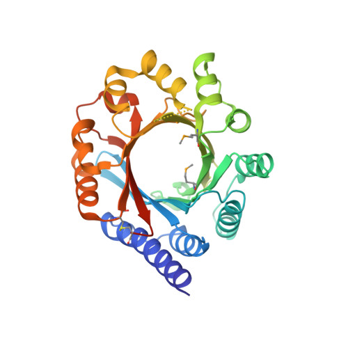
Structure of Patf from Prochloron Didemni.

Bent, A.F.Koehnke, J.Houssen, W.E.Smith, M.C.M.Jaspars, M.Naismith, J.H.

(2013) Acta Crystallogr Sect F Struct Biol Cryst Commun 69: 618

  • DOI: https://doi.org/10.1107/S1744309113012931
  • Primary Citation of Related Structures:  
    4BG2

  • PubMed Abstract: 

    Patellamides are macrocyclic peptides with potent biological effects and are a subset of the cyanobactins. Cyanobactins are natural products that are produced by a series of enzymatic transformations and a common modification is the addition of a prenyl group. Puzzlingly, the pathway for patellamides in Prochloron didemni contains a gene, patF, with homology to prenylases, but patellamides are not themselves prenylated. The structure of the protein PatF was cloned, expressed, purified and determined. Prenylase activity could not be demonstrated for the protein, and examination of the structure revealed changes in side-chain identity at the active site. It is suggested that these changes have inactivated the protein. Attempts to mutate these residues led to unfolded protein.


  • Organizational Affiliation

    Biomedical Sciences Research Complex, University of St Andrews, North Haugh, St Andrews, Fife KY16 9ST, Scotland.


Macromolecules
Find similar proteins by:  (by identity cutoff)  |  3D Structure
Entity ID: 1
MoleculeChains Sequence LengthOrganismDetailsImage
PATF
A, B
321Prochloron didemniMutation(s): 0 
UniProt
Find proteins for Q52QJ2 (Prochloron didemni)
Explore Q52QJ2 
Go to UniProtKB:  Q52QJ2
Entity Groups  
Sequence Clusters30% Identity50% Identity70% Identity90% Identity95% Identity100% Identity
UniProt GroupQ52QJ2
Sequence Annotations
Expand
  • Reference Sequence
Small Molecules
Modified Residues  1 Unique
IDChains TypeFormula2D DiagramParent
MSE
Query on MSE
A, B
L-PEPTIDE LINKINGC5 H11 N O2 SeMET
Experimental Data & Validation

Experimental Data

  • Method: X-RAY DIFFRACTION
  • Resolution: 2.13 Å
  • R-Value Free: 0.221 
  • R-Value Work: 0.190 
  • R-Value Observed: 0.192 
  • Space Group: P 1 21 1
Unit Cell:
Length ( Å )Angle ( ˚ )
a = 47.08α = 90
b = 135.8β = 118.54
c = 48.738γ = 90
Software Package:
Software NamePurpose
REFMACrefinement
XDSdata reduction
SCALAdata scaling
PHENIXphasing

Structure Validation

View Full Validation Report


Entry History 

Deposition Data

Revision History  (Full details and data files)

  • Version 1.0: 2013-04-03
    Type: Initial release
  • Version 1.1: 2013-06-12
    Changes: Database references
  • Version 1.2: 2013-11-06
    Changes: Structure summary