Keyhole Limpet Hemocyanin: 9-A Cryoem Structure and Molecular Model of the Klh1 Didecamer Reveal the Interfaces and Intricate Topology of the 160 Functional Units.
Gatsogiannis, C., Markl, J.(2009) J Mol Biol 385: 963
- PubMed: 19013468
- DOI: https://doi.org/10.1016/j.jmb.2008.10.080
- Primary Citation of Related Structures:
4BED - PubMed Abstract:
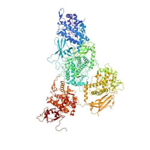
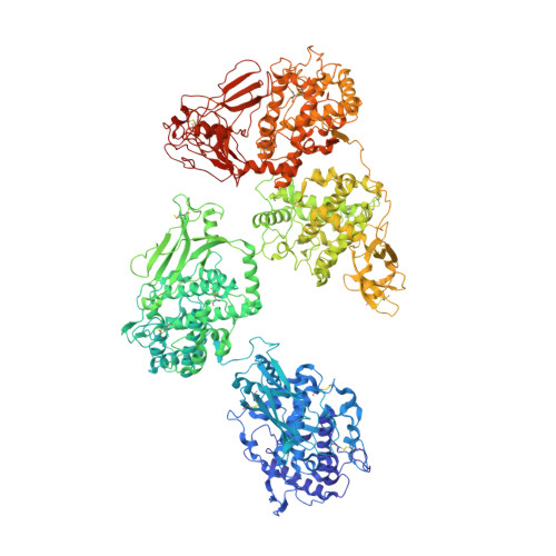
Hemocyanins are blue copper-containing respiratory proteins in the hemolymph of many arthropods and molluscs. Molluscan hemocyanins are decamers, didecamers, or multidecamers of a 340- to 400-kDa polypeptide subunit containing seven or eight globular functional units (FUs; FU-a to FU-h), each with an oxygen-binding site. The decamers are short 35-nm hollow cylinders, with their lumen narrowed by a collar complex. Our recently published 9-A cryo-electron microscopy/crystal structure hybrid model of a 3.4-MDa cephalopod hemocyanin decamer [Nautilus pompilius hemocyanin (NpH)] revealed the pathway of the seven-FU subunit (340 kDa), 15 types of inter-FU interface, and an asymmetric collar consisting of five "arcs" (FU-g pairs). We now present a comparable hybrid model of an 8-MDa gastropod hemocyanin didecamer assembled from two asymmetric decamers [isoform keyhole limpet hemocyanin (KLH) 1 of the established immunogen KLH]. Compared to NpH, the KLH1 subunit (400 kDa) is C-terminally elongated by FU-h, which is further extended by a unique tail domain. We have found that the wall-and-arc structure of the KLH1 decamer is very similar to that of NpH. We have traced the subunit pathway and how it continues from KLH1-g to KLH1-h to form an annulus of five "slabs" (FU-h pairs) at one cylinder edge. The 15 types of inter-FU interface detected in NpH are also present in KLH1. Moreover, we have identified one arc/slab interface, two slab/slab interfaces, five slab/wall interfaces, and four decamer/decamer interfaces. The 27 interfaces are described on the basis of two subunit conformers, yielding an asymmetric homodimer. Six protrusions from the cryo-electron microscopy structure per subunit are associated with putative attachment sites for N-linked glycans, indicating a total of 120 sugar trees in KLH1. Also, putative binding sites for divalent cations have been detected. In conclusion, the present 9-A data on KLH1 confirm and substantially broaden our recent analysis of the smaller cephalopod hemocyanin and essentially solve the gastropod hemocyanin structure.
Organizational Affiliation:
Institute of Zoology, Johannes Gutenberg University, D-55099 Mainz, Germany.