4AY8

SeMet-derivative of a methyltransferase from M. mazei


Experimental Data Snapshot

  • Method: X-RAY DIFFRACTION
  • Resolution: 2.10 Å
  • R-Value Free: 0.177 
  • R-Value Work: 0.146 
  • R-Value Observed: 0.148 

wwPDB Validation   3D Report Full Report


This is version 1.2 of the entry. See complete history


Literature

Structure of the Corrinoid:Coenzyme M Methyltransferase Mtaa from Methanosarcina Mazei

Hoeppner, A.Thomas, F.Rueppel, A.Hensel, R.Blankenfeldt, W.Bayer, P.Faust, A.

(2012) Acta Crystallogr D Biol Crystallogr 68: 1549

  • DOI: https://doi.org/10.1107/S090744491203853X
  • Primary Citation of Related Structures:  
    4AY7, 4AY8

  • PubMed Abstract: 

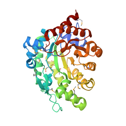
    The zinc-containing corrinoid:coenzyme M methyltransferase MtaA is part of the methanol-coenzyme M-methyltransferase complex of Methanosarcina mazei. The whole complex consists of three subunits: MtaA, MtaB and MtaC. The MtaB-MtaC complex catalyses the cleavage of methanol (bound to MtaB) and the transfer of the methyl group onto the cobalt of cob(I)alamin (bound to MtaC). The MtaA-MtaC complex catalyses methyl transfer from methyl-cob(III)alamin (bound to MtaC) to coenzyme M (bound to MtaA). The crystal structure of the MtaB-MtaC complex from M. barkeri has previously been determined. Here, the crystal structures of MtaA from M. mazei in a substrate-free but Zn(2+)-bound state and in complex with Zn(2+) and coenzyme M (HS-CoM) are reported at resolutions of 1.8 and 2.1 Å, respectively. A search for homologous proteins revealed that MtaA exhibits 23% sequence identity to human uroporphyrinogen III decarboxylase, which has also the highest structural similarity (r.m.s.d. of 2.03 Å for 306 aligned amino acids). The main structural feature of MtaA is a TIM-barrel-like fold, which is also found in all other zinc enzymes that catalyse thiol-group alkylation. The active site of MtaA is situated at the narrow bottom of a funnel such that the thiolate group of HS-CoM points towards the Zn(2+) ion. The Zn(2+) ion in the active site of MtaA is coordinated tetrahedrally via His240, Cys242 and Cys319. In the substrate-free form the fourth ligand is Glu263. Binding of HS-CoM leads to exchange of the O-ligand of Glu263 for the S-ligand of HS-CoM with inversion of the zinc geometry. The interface between MtaA and MtaC for transfer of the methyl group from MtaC-bound methylcobalamin is most likely to be formed by the core complex of MtaB-MtaC and the N-terminal segment (a long loop containing three α-helices and a β-hairpin) of MtaA, which is not part of the TIM-barrel core structure of MtaA.


  • Organizational Affiliation

    Structural and Medicinal Biochemistry, Centre of Medical Biotechnology (ZMB), University of Duisburg-Essen, Campus Essen, 45117 Essen, Germany.


Macromolecules
Find similar proteins by:  (by identity cutoff)  |  3D Structure
Entity ID: 1
MoleculeChains Sequence LengthOrganismDetailsImage
METHYLCOBALAMIN\: COENZYME M METHYLTRANSFERASE
A, B
348Methanosarcina mazeiMutation(s): 0 
EC: 2.1.1.247
UniProt
Find proteins for Q8PXZ6 (Methanosarcina mazei (strain ATCC BAA-159 / DSM 3647 / Goe1 / Go1 / JCM 11833 / OCM 88))
Explore Q8PXZ6 
Go to UniProtKB:  Q8PXZ6
Entity Groups  
Sequence Clusters30% Identity50% Identity70% Identity90% Identity95% Identity100% Identity
UniProt GroupQ8PXZ6
Sequence Annotations
Expand
  • Reference Sequence
Small Molecules
Ligands 4 Unique
IDChains Name / Formula / InChI Key2D Diagram3D Interactions
COM
Query on COM

Download Ideal Coordinates CCD File 
C [auth A],
F [auth B]
1-THIOETHANESULFONIC ACID
C2 H6 O3 S2
ZNEWHQLOPFWXOF-UHFFFAOYSA-N
MRD
Query on MRD

Download Ideal Coordinates CCD File 
E [auth A](4R)-2-METHYLPENTANE-2,4-DIOL
C6 H14 O2
SVTBMSDMJJWYQN-RXMQYKEDSA-N
GOL
Query on GOL

Download Ideal Coordinates CCD File 
H [auth B],
I [auth B]
GLYCEROL
C3 H8 O3
PEDCQBHIVMGVHV-UHFFFAOYSA-N
ZN
Query on ZN

Download Ideal Coordinates CCD File 
D [auth A],
G [auth B]
ZINC ION
Zn
PTFCDOFLOPIGGS-UHFFFAOYSA-N
Modified Residues  2 Unique
IDChains TypeFormula2D DiagramParent
CSD
Query on CSD
A, B
L-PEPTIDE LINKINGC3 H7 N O4 SCYS
MSE
Query on MSE
A, B
L-PEPTIDE LINKINGC5 H11 N O2 SeMET
Experimental Data & Validation

Experimental Data

  • Method: X-RAY DIFFRACTION
  • Resolution: 2.10 Å
  • R-Value Free: 0.177 
  • R-Value Work: 0.146 
  • R-Value Observed: 0.148 
  • Space Group: P 32
Unit Cell:
Length ( Å )Angle ( ˚ )
a = 125.21α = 90
b = 125.21β = 90
c = 38.88γ = 120
Software Package:
Software NamePurpose
REFMACrefinement
XDSdata reduction
SCALAdata scaling
SHELXphasing

Structure Validation

View Full Validation Report


Entry History 

Deposition Data

Revision History  (Full details and data files)

  • Version 1.0: 2012-10-31
    Type: Initial release
  • Version 1.1: 2012-11-07
    Changes: Database references
  • Version 1.2: 2014-11-05
    Changes: Atomic model, Derived calculations, Non-polymer description, Other, Structure summary