4AW9

Crystal structure of active legumain in complex with YVAD-CMK


Experimental Data Snapshot

  • Method: X-RAY DIFFRACTION
  • Resolution: 2.20 Å
  • R-Value Free: 0.207 
  • R-Value Work: 0.180 
  • R-Value Observed: 0.182 

wwPDB Validation   3D Report Full Report


Ligand Structure Quality Assessment 


This is version 1.3 of the entry. See complete history


Literature

Mechanistic and Structural Studies on Legumain Explain its Zymogenicity, Distinct Activation Pathways, and Regulation

Dall, E.Brandstetter, H.

(2013) Proc Natl Acad Sci U S A 110: 10940

  • DOI: https://doi.org/10.1073/pnas.1300686110
  • Primary Citation of Related Structures:  
    4AW9, 4AWA, 4AWB, 4FGU

  • PubMed Abstract: 

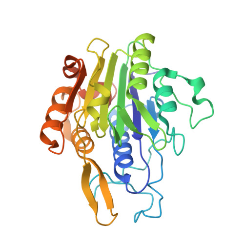
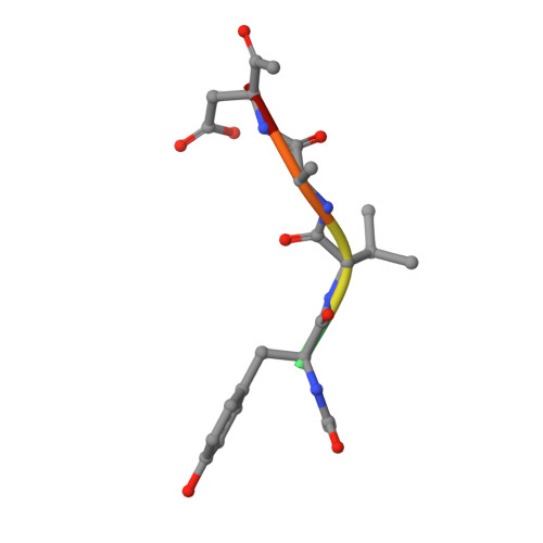
    The cysteine protease legumain plays important functions in immunity and cancer at different cellular locations, some of which appeared conflicting with its proteolytic activity and stability. Here, we report crystal structures of legumain in the zymogenic and fully activated form in complex with different substrate analogs. We show that the eponymous asparagine-specific endopeptidase activity is electrostatically generated by pH shift. Completely unexpectedly, the structure points toward a hidden carboxypeptidase activity that develops upon proteolytic activation with the release of an activation peptide. These activation routes reconcile the enigmatic pH stability of legumain, e.g., lysosomal, nuclear, and extracellular activities with relevance in immunology and cancer. Substrate access and turnover is controlled by selective protonation of the S1 pocket (KM) and the catalytic nucleophile (kcat), respectively. The multibranched and context-dependent activation process of legumain illustrates how proteases can act not only as signal transducers but as decision makers.


  • Organizational Affiliation

    Department of Molecular Biology, University of Salzburg, A-5020 Salzburg, Austria.


Macromolecules
Find similar proteins by:  (by identity cutoff)  |  3D Structure
Entity ID: 1
MoleculeChains Sequence LengthOrganismDetailsImage
LEGUMAIN284Homo sapiensMutation(s): 1 
EC: 3.4.22.34
UniProt & NIH Common Fund Data Resources
Find proteins for Q99538 (Homo sapiens)
Explore Q99538 
Go to UniProtKB:  Q99538
PHAROS:  Q99538
GTEx:  ENSG00000100600 
Entity Groups  
Sequence Clusters30% Identity50% Identity70% Identity90% Identity95% Identity100% Identity
UniProt GroupQ99538
Sequence Annotations
Expand
  • Reference Sequence

Find similar proteins by:  Sequence   |   3D Structure  

Entity ID: 2
MoleculeChains Sequence LengthOrganismDetailsImage
ACE-TYR-VAL-ALA-ASP-CHLOROMETHYLKETONEB [auth I]6synthetic constructMutation(s): 0 
Sequence Annotations
Expand
  • Reference Sequence
Biologically Interesting Molecules (External Reference) 1 Unique
Experimental Data & Validation

Experimental Data

  • Method: X-RAY DIFFRACTION
  • Resolution: 2.20 Å
  • R-Value Free: 0.207 
  • R-Value Work: 0.180 
  • R-Value Observed: 0.182 
  • Space Group: P 42
Unit Cell:
Length ( Å )Angle ( ˚ )
a = 64.14α = 90
b = 64.14β = 90
c = 78.95γ = 90
Software Package:
Software NamePurpose
REFMACrefinement
iMOSFLMdata reduction
SCALAdata scaling

Structure Validation

View Full Validation Report


Ligand Structure Quality Assessment 


Entry History 

Deposition Data

Revision History  (Full details and data files)

  • Version 1.0: 2013-06-26
    Type: Initial release
  • Version 1.1: 2013-07-17
    Changes: Database references
  • Version 1.2: 2013-10-02
    Changes: Other
  • Version 1.3: 2020-07-29
    Type: Remediation
    Reason: Carbohydrate remediation
    Changes: Data collection, Derived calculations, Other, Structure summary