4AQJ

Structure of human S100A7 D24G bound to zinc and calcium


Experimental Data Snapshot

  • Method: X-RAY DIFFRACTION
  • Resolution: 1.60 Å
  • R-Value Free: 0.225 
  • R-Value Work: 0.189 
  • R-Value Observed: 0.191 

wwPDB Validation   3D Report Full Report


This is version 1.0 of the entry. See complete history


Literature

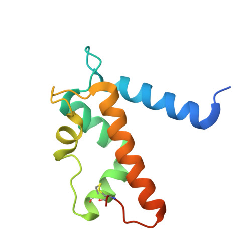
Structural Characterization of S100A15 Reveals a Novel Zinc Coordination Site Among S100 Proteins and Altered Surface Chemistry with Functional Implications for Receptor Binding.

Murray, J.I.Tonkin, M.L.Whiting, A.L.Peng, F.Farnell, B.Cullen, J.T.Hof, F.Boulanger, M.J.

(2012) BMC Struct Biol 12: 16

  • DOI: https://doi.org/10.1186/1472-6807-12-16
  • Primary Citation of Related Structures:  
    4AQI, 4AQJ

  • PubMed Abstract: 

    S100 proteins are a family of small, EF-hand containing calcium-binding signaling proteins that are implicated in many cancers. While the majority of human S100 proteins share 25-65% sequence similarity, S100A7 and its recently identified paralog, S100A15, display 93% sequence identity. Intriguingly, however, S100A7 and S100A15 serve distinct roles in inflammatory skin disease; S100A7 signals through the receptor for advanced glycation products (RAGE) in a zinc-dependent manner, while S100A15 signals through a yet unidentified G-protein coupled receptor in a zinc-independent manner. Of the seven divergent residues that differentiate S100A7 and S100A15, four cluster in a zinc-binding region and the remaining three localize to a predicted receptor-binding surface.


  • Organizational Affiliation

    Department of Chemistry, University of Victoria, PO Box 3065STN CSC, Victoria, BC V8W 3P6, Canada.


Macromolecules
Find similar proteins by:  (by identity cutoff)  |  3D Structure
Entity ID: 1
MoleculeChains Sequence LengthOrganismDetailsImage
PROTEIN S100-A7109Homo sapiensMutation(s): 2 
UniProt & NIH Common Fund Data Resources
Find proteins for P31151 (Homo sapiens)
Explore P31151 
Go to UniProtKB:  P31151
PHAROS:  P31151
GTEx:  ENSG00000143556 
Entity Groups  
Sequence Clusters30% Identity50% Identity70% Identity90% Identity95% Identity100% Identity
UniProt GroupP31151
Sequence Annotations
Expand
  • Reference Sequence
Experimental Data & Validation

Experimental Data

  • Method: X-RAY DIFFRACTION
  • Resolution: 1.60 Å
  • R-Value Free: 0.225 
  • R-Value Work: 0.189 
  • R-Value Observed: 0.191 
  • Space Group: P 43 21 2
Unit Cell:
Length ( Å )Angle ( ˚ )
a = 51.48α = 90
b = 51.48β = 90
c = 117.23γ = 90
Software Package:
Software NamePurpose
REFMACrefinement
d*TREKdata reduction
MOLREPphasing

Structure Validation

View Full Validation Report


Entry History 

Deposition Data

Revision History  (Full details and data files)

  • Version 1.0: 2012-10-17
    Type: Initial release