4ZZB

The GLIC pentameric Ligand-Gated Ion Channel Locally-closed form complexed to xenon


Experimental Data Snapshot

  • Method: X-RAY DIFFRACTION
  • Resolution: 3.40 Å
  • R-Value Free: 0.223 
  • R-Value Work: 0.216 
  • R-Value Observed: 0.217 

wwPDB Validation   3D Report Full Report


Ligand Structure Quality Assessment 


This is version 1.2 of the entry. See complete history


Literature

Structural Basis for Xenon Inhibition in a Cationic Pentameric Ligand-Gated Ion Channel.

Sauguet, L.Fourati, Z.Prange, T.Delarue, M.Colloc'h, N.

(2016) PLoS One 11: e0149795-e0149795

  • DOI: https://doi.org/10.1371/journal.pone.0149795
  • Primary Citation of Related Structures:  
    4ZZB, 4ZZC

  • PubMed Abstract: 

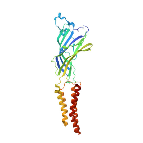
    GLIC receptor is a bacterial pentameric ligand-gated ion channel whose action is inhibited by xenon. Xenon has been used in clinical practice as a potent gaseous anaesthetic for decades, but the molecular mechanism of interactions with its integral membrane receptor targets remains poorly understood. Here we characterize by X-ray crystallography the xenon-binding sites within both the open and "locally-closed" (inactive) conformations of GLIC. Major binding sites of xenon, which differ between the two conformations, were identified in three distinct regions that all belong to the trans-membrane domain of GLIC: 1) in an intra-subunit cavity, 2) at the interface between adjacent subunits, and 3) in the pore. The pore site is unique to the locally-closed form where the binding of xenon effectively seals the channel. A putative mechanism of the inhibition of GLIC by xenon is proposed, which might be extended to other pentameric cationic ligand-gated ion channels.


  • Organizational Affiliation

    Unité de Dynamique Structurale des Macromolécules (UMR 3528 CNRS) Institut Pasteur, Paris, France.


Macromolecules
Find similar proteins by:  (by identity cutoff)  |  3D Structure
Entity ID: 1
MoleculeChains Sequence LengthOrganismDetailsImage
Proton-gated ion channel
A, B, C, D, E
317Gloeobacter violaceus PCC 8105Mutation(s): 3 
Gene Names: glvIglr4197
Membrane Entity: Yes 
UniProt
Find proteins for Q7NDN8 (Gloeobacter violaceus (strain ATCC 29082 / PCC 7421))
Explore Q7NDN8 
Go to UniProtKB:  Q7NDN8
Entity Groups  
Sequence Clusters30% Identity50% Identity70% Identity90% Identity95% Identity100% Identity
UniProt GroupQ7NDN8
Sequence Annotations
Expand
  • Reference Sequence
Small Molecules
Ligands 5 Unique
IDChains Name / Formula / InChI Key2D Diagram3D Interactions
LMT
Query on LMT

Download Ideal Coordinates CCD File 
F [auth A]DODECYL-BETA-D-MALTOSIDE
C24 H46 O11
NLEBIOOXCVAHBD-QKMCSOCLSA-N
XE
Query on XE

Download Ideal Coordinates CCD File 
AA [auth C]
BA [auth C]
CA [auth C]
DA [auth C]
HA [auth D]
AA [auth C],
BA [auth C],
CA [auth C],
DA [auth C],
HA [auth D],
IA [auth D],
J [auth A],
JA [auth D],
K [auth A],
KA [auth D],
L [auth A],
M [auth A],
N [auth A],
O [auth A],
OA [auth E],
PA [auth E],
QA [auth E],
S [auth B],
T [auth B],
U [auth B],
V [auth B]
XENON
Xe
FHNFHKCVQCLJFQ-UHFFFAOYSA-N
ACT
Query on ACT

Download Ideal Coordinates CCD File 
GA [auth D]
I [auth A]
NA [auth E]
R [auth B]
W [auth C]
GA [auth D],
I [auth A],
NA [auth E],
R [auth B],
W [auth C],
Z [auth C]
ACETATE ION
C2 H3 O2
QTBSBXVTEAMEQO-UHFFFAOYSA-M
CL
Query on CL

Download Ideal Coordinates CCD File 
EA [auth D],
G [auth A],
LA [auth E],
P [auth B],
X [auth C]
CHLORIDE ION
Cl
VEXZGXHMUGYJMC-UHFFFAOYSA-M
NA
Query on NA

Download Ideal Coordinates CCD File 
FA [auth D],
H [auth A],
MA [auth E],
Q [auth B],
Y [auth C]
SODIUM ION
Na
FKNQFGJONOIPTF-UHFFFAOYSA-N
Experimental Data & Validation

Experimental Data

  • Method: X-RAY DIFFRACTION
  • Resolution: 3.40 Å
  • R-Value Free: 0.223 
  • R-Value Work: 0.216 
  • R-Value Observed: 0.217 
  • Space Group: C 1 2 1
Unit Cell:
Length ( Å )Angle ( ˚ )
a = 176.966α = 90
b = 130.528β = 100.33
c = 157.526γ = 90
Software Package:
Software NamePurpose
BUSTERrefinement
XDSdata reduction
SCALAdata scaling
REFMACphasing

Structure Validation

View Full Validation Report


Ligand Structure Quality Assessment 


Entry History 

Deposition Data

Revision History  (Full details and data files)

  • Version 1.0: 2016-03-02
    Type: Initial release
  • Version 1.1: 2016-03-09
    Changes: Database references
  • Version 1.2: 2018-11-21
    Changes: Data collection, Source and taxonomy