4ZYR

Crystal structure of E. coli Lactose permease G46W/G262W bound to p-nitrophenyl alpha-D-galactopyranoside (alpha-NPG)


Experimental Data Snapshot

  • Method: X-RAY DIFFRACTION
  • Resolution: 3.31 Å
  • R-Value Free: 0.278 
  • R-Value Work: 0.259 
  • R-Value Observed: 0.260 

wwPDB Validation   3D Report Full Report


Ligand Structure Quality Assessment 


This is version 1.6 of the entry. See complete history


Literature

Structure of LacY with an alpha-substituted galactoside: Connecting the binding site to the protonation site.

Kumar, H.Finer-Moore, J.S.Kaback, H.R.Stroud, R.M.

(2015) Proc Natl Acad Sci U S A 112: 9004-9009

  • DOI: https://doi.org/10.1073/pnas.1509854112
  • Primary Citation of Related Structures:  
    4ZYR

  • PubMed Abstract: 

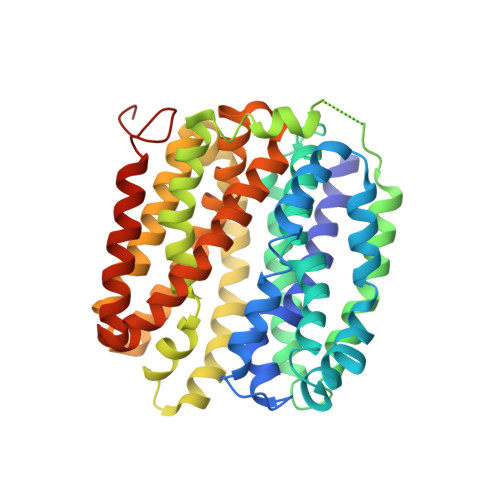
    The X-ray crystal structure of a conformationally constrained mutant of the Escherichia coli lactose permease (the LacY double-Trp mutant Gly-46→Trp/Gly-262→Trp) with bound p-nitrophenyl-α-d-galactopyranoside (α-NPG), a high-affinity lactose analog, is described. With the exception of Glu-126 (helix IV), side chains Trp-151 (helix V), Glu-269 (helix VIII), Arg-144 (helix V), His-322 (helix X), and Asn-272 (helix VIII) interact directly with the galactopyranosyl ring of α-NPG to provide specificity, as indicated by biochemical studies and shown directly by X-ray crystallography. In contrast, Phe-20, Met-23, and Phe-27 (helix I) are within van der Waals distance of the benzyl moiety of the analog and thereby increase binding affinity nonspecifically. Thus, the specificity of LacY for sugar is determined solely by side-chain interactions with the galactopyranosyl ring, whereas affinity is increased by nonspecific hydrophobic interactions with the anomeric substituent.


  • Organizational Affiliation

    Department of Biochemistry and Biophysics, University of California, San Francisco, CA 94158;


Macromolecules
Find similar proteins by:  (by identity cutoff)  |  3D Structure
Entity ID: 1
MoleculeChains Sequence LengthOrganismDetailsImage
Lactose permease
A, B
417Escherichia coli K-12Mutation(s): 2 
Gene Names: lacYb0343JW0334
Membrane Entity: Yes 
UniProt
Find proteins for P02920 (Escherichia coli (strain K12))
Explore P02920 
Go to UniProtKB:  P02920
Entity Groups  
Sequence Clusters30% Identity50% Identity70% Identity90% Identity95% Identity100% Identity
UniProt GroupP02920
Sequence Annotations
Expand
  • Reference Sequence
Experimental Data & Validation

Experimental Data

  • Method: X-RAY DIFFRACTION
  • Resolution: 3.31 Å
  • R-Value Free: 0.278 
  • R-Value Work: 0.259 
  • R-Value Observed: 0.260 
  • Space Group: C 2 2 21
Unit Cell:
Length ( Å )Angle ( ˚ )
a = 103.15α = 90
b = 121.99β = 90
c = 266.09γ = 90
Software Package:
Software NamePurpose
PHENIXrefinement
XDSdata reduction
XDSdata scaling
PHASERphasing

Structure Validation

View Full Validation Report


Ligand Structure Quality Assessment 


Entry History & Funding Information

Deposition Data


Funding OrganizationLocationGrant Number
National Institutes of Health/National Institute of General Medical Sciences (NIH/NIGMS)United StatesR37GM024485
National Institutes of Health/National Institute of Diabetes and Digestive and Kidney Disease (NIH/NIDDK)United StatesDK069463
National Science Foundation (NSF, United States)United StatesMCB-1129551

Revision History  (Full details and data files)

  • Version 1.0: 2015-07-29
    Type: Initial release
  • Version 1.1: 2015-08-05
    Changes: Database references
  • Version 1.2: 2015-08-12
    Changes: Derived calculations
  • Version 1.3: 2017-09-20
    Changes: Author supporting evidence, Derived calculations
  • Version 1.4: 2019-11-27
    Changes: Author supporting evidence
  • Version 1.5: 2020-07-29
    Type: Remediation
    Reason: Carbohydrate remediation
    Changes: Data collection, Derived calculations, Structure summary
  • Version 1.6: 2023-09-27
    Changes: Data collection, Database references, Refinement description, Structure summary