4ZKL

Crystal structure of human histidine triad nucleotide-binding protein 1 (hHINT1) complexed with JB419 (AP4A analog)


Experimental Data Snapshot

  • Method: X-RAY DIFFRACTION
  • Resolution: 2.34 Å
  • R-Value Free: 0.239 
  • R-Value Work: 0.186 
  • R-Value Observed: 0.189 

wwPDB Validation   3D Report Full Report


Ligand Structure Quality Assessment 


This is version 1.3 of the entry. See complete history


Literature

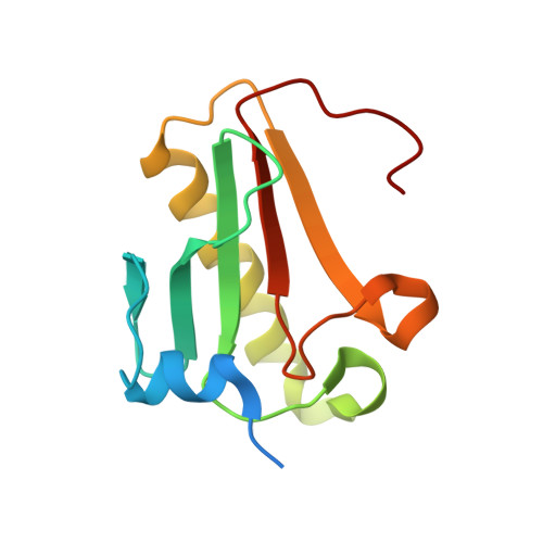
Crystallographic studies of the complex of human HINT1 protein with a non-hydrolyzable analog of Ap4A.

Dolot, R.Kaczmarek, R.Seda, A.Krakowiak, A.Baraniak, J.Nawrot, B.

(2016) Int J Biol Macromol 87: 62-69

  • DOI: https://doi.org/10.1016/j.ijbiomac.2016.02.047
  • Primary Citation of Related Structures:  
    4ZKL, 4ZKV

  • PubMed Abstract: 

    Histidine triad nucleotide-binding protein 1 (HINT1) represents the most ancient and widespread branch in the histidine triad proteins superfamily. HINT1 plays an important role in various biological processes, and it has been found in many species. Here, we report the first structure (at a 2.34Å resolution) of a complex of human HINT1 with a non-hydrolyzable analog of an Ap4A dinucleotide, containing bis-phosphorothioated glycerol mimicking a polyphosphate chain, obtained from a primitive monoclinic space group P21 crystal. In addition, the apo form of hHINT1 at the space group P21 refined to 1.92Å is reported for comparative studies.


  • Organizational Affiliation

    Department of Bioorganic Chemistry, Centre of Molecular and Macromolecular Studies, Polish Academy of Sciences, Sienkiewicza 112, 90-363 Łódź, Poland. Electronic address: rdolot@cbmm.lodz.pl.


Macromolecules
Find similar proteins by:  (by identity cutoff)  |  3D Structure
Entity ID: 1
MoleculeChains Sequence LengthOrganismDetailsImage
Histidine triad nucleotide-binding protein 1
A, B, C, D
126Homo sapiensMutation(s): 0 
Gene Names: HINT1HINTPKCI1PRKCNH1
EC: 3
UniProt & NIH Common Fund Data Resources
Find proteins for P49773 (Homo sapiens)
Explore P49773 
Go to UniProtKB:  P49773
PHAROS:  P49773
GTEx:  ENSG00000169567 
Entity Groups  
Sequence Clusters30% Identity50% Identity70% Identity90% Identity95% Identity100% Identity
UniProt GroupP49773
Sequence Annotations
Expand
  • Reference Sequence
Small Molecules
Ligands 2 Unique
IDChains Name / Formula / InChI Key2D Diagram3D Interactions
JB6
Query on JB6

Download Ideal Coordinates CCD File 
F [auth B](2R,3R,4S,5R)-2-(6-aminopurin-9-yl)-5-[2-[[(2S)-3-[[(2R,3S,4R,5R)-5-(6-aminopurin-9-yl)-3,4-bis(oxidanyl)oxolan-2-yl]methoxy-sulfanyl-phosphoryl]oxy-2-oxidanyl-propoxy]-sulfanyl-phosphoryl]oxyethyl]oxolane-3,4-diol
C24 H34 N10 O13 P2 S2
OABPMBSLNAAXBR-CZUYOEIMSA-N
AMP
Query on AMP

Download Ideal Coordinates CCD File 
E [auth B],
G [auth D]
ADENOSINE MONOPHOSPHATE
C10 H14 N5 O7 P
UDMBCSSLTHHNCD-KQYNXXCUSA-N
Experimental Data & Validation

Experimental Data

  • Method: X-RAY DIFFRACTION
  • Resolution: 2.34 Å
  • R-Value Free: 0.239 
  • R-Value Work: 0.186 
  • R-Value Observed: 0.189 
  • Space Group: P 1 21 1
Unit Cell:
Length ( Å )Angle ( ˚ )
a = 63.714α = 90
b = 46.419β = 97.41
c = 103.413γ = 90
Software Package:
Software NamePurpose
REFMACrefinement
XDSdata reduction
Aimlessdata scaling
MOLREPphasing

Structure Validation

View Full Validation Report


Ligand Structure Quality Assessment 


Entry History 

Deposition Data

Revision History  (Full details and data files)

  • Version 1.0: 2016-03-02
    Type: Initial release
  • Version 1.1: 2016-03-09
    Changes: Database references
  • Version 1.2: 2018-03-07
    Changes: Data collection
  • Version 1.3: 2024-01-10
    Changes: Data collection, Database references, Refinement description