


Crystal structure of aminoglycoside acetyltransferase AAC(3)-Ib
Stogios, P.J.To be published.
Experimental Data Snapshot
wwPDB Validation 3D Report Full Report

Entity ID: 1 | |||||
|---|---|---|---|---|---|
| Molecule | Chains | Sequence Length | Organism | Details | Image |
| Aminoglycoside 3'-N-acetyltransferase | 182 | Pseudomonas aeruginosa | Mutation(s): 0 Gene Names: aac(3)-Ib | 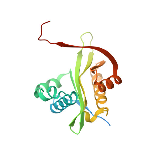 | |
UniProt | |||||
Find proteins for Q51407 (Pseudomonas aeruginosa) Explore Q51407 Go to UniProtKB: Q51407 | |||||
Entity Groups | |||||
| Sequence Clusters | 30% Identity50% Identity70% Identity90% Identity95% Identity100% Identity | ||||
| UniProt Group | Q51407 | ||||
Sequence AnnotationsExpand | |||||
| |||||
| Ligands 1 Unique | |||||
|---|---|---|---|---|---|
| ID | Chains | Name / Formula / InChI Key | 2D Diagram | 3D Interactions | |
| SO4 Query on SO4 | C [auth A] D [auth A] E [auth A] F [auth A] G [auth B] | SULFATE ION O4 S QAOWNCQODCNURD-UHFFFAOYSA-L |  | ||
| Length ( Å ) | Angle ( ˚ ) |
|---|---|
| a = 77.024 | α = 90 |
| b = 77.024 | β = 90 |
| c = 128.66 | γ = 90 |
| Software Name | Purpose |
|---|---|
| PHENIX | refinement |
| HKL-3000 | data reduction |
| HKL-3000 | data scaling |
| PHENIX | phasing |
| Coot | model building |
RCSB PDB (citation) is hosted by
RCSB PDB is a member of the

