4YAO

Reduced CYPOR mutant - G143del


Experimental Data Snapshot

  • Method: X-RAY DIFFRACTION
  • Resolution: 2.50 Å
  • R-Value Free: 0.298 
  • R-Value Work: 0.234 
  • R-Value Observed: 0.234 

wwPDB Validation   3D Report Full Report


Ligand Structure Quality Assessment 


This is version 1.6 of the entry. See complete history


Literature

Mutants of Cytochrome P450 Reductase Lacking Either Gly-141 or Gly-143 Destabilize Its FMN Semiquinone.

Rwere, F.Xia, C.Im, S.Haque, M.M.Stuehr, D.J.Waskell, L.Kim, J.J.

(2016) J Biol Chem 291: 14639-14661

  • DOI: https://doi.org/10.1074/jbc.M116.724625
  • Primary Citation of Related Structures:  
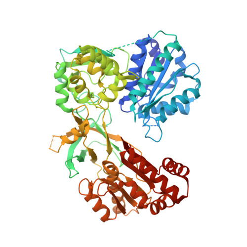
    4Y7C, 4Y9R, 4Y9U, 4YAF, 4YAL, 4YAO, 4YAU, 4YAW

  • PubMed Abstract: 

    NADPH-cytochrome P450 oxidoreductase transfers electrons from NADPH to cytochromes P450 via its FAD and FMN. To understand the biochemical and structural basis of electron transfer from FMN-hydroquinone to its partners, three deletion mutants in a conserved loop near the FMN were characterized. Comparison of oxidized and reduced wild type and mutant structures reveals that the basis for the air stability of the neutral blue semiquinone is protonation of the flavin N5 and strong H-bond formation with the Gly-141 carbonyl. The ΔGly-143 protein had moderately decreased activity with cytochrome P450 and cytochrome c It formed a flexible loop, which transiently interacts with the flavin N5, resulting in the generation of both an unstable neutral blue semiquinone and hydroquinone. The ΔGly-141 and ΔG141/E142N mutants were inactive with cytochrome P450 but fully active in reducing cytochrome c In the ΔGly-141 mutants, the backbone amide of Glu/Asn-142 forms an H-bond to the N5 of the oxidized flavin, which leads to formation of an unstable red anionic semiquinone with a more negative potential than the hydroquinone. The semiquinone of ΔG141/E142N was slightly more stable than that of ΔGly-141, consistent with its crystallographically demonstrated more rigid loop. Nonetheless, both ΔGly-141 red semiquinones were less stable than those of the corresponding loop in cytochrome P450 BM3 and the neuronal NOS mutant (ΔGly-810). Our results indicate that the catalytic activity of cytochrome P450 oxidoreductase is a function of the length, sequence, and flexibility of the 140s loop and illustrate the sophisticated variety of biochemical mechanisms employed in fine-tuning its redox properties and function.


  • Organizational Affiliation

    From the Department of Anesthesiology, University of Michigan and Veterans Affairs Medical Center, Ann Arbor, Michigan 48105.


Macromolecules
Find similar proteins by:  (by identity cutoff)  |  3D Structure
Entity ID: 1
MoleculeChains Sequence LengthOrganismDetailsImage
NADPH--cytochrome P450 reductase
A, B
621Rattus norvegicusMutation(s): 0 
Gene Names: Por
EC: 1.6.2.4
UniProt
Find proteins for P00388 (Rattus norvegicus)
Explore P00388 
Go to UniProtKB:  P00388
Entity Groups  
Sequence Clusters30% Identity50% Identity70% Identity90% Identity95% Identity100% Identity
UniProt GroupP00388
Sequence Annotations
Expand
  • Reference Sequence
Small Molecules
Ligands 4 Unique
IDChains Name / Formula / InChI Key2D Diagram3D Interactions
FAD
Query on FAD

Download Ideal Coordinates CCD File 
D [auth A],
I [auth B]
FLAVIN-ADENINE DINUCLEOTIDE
C27 H33 N9 O15 P2
VWWQXMAJTJZDQX-UYBVJOGSSA-N
FMN
Query on FMN

Download Ideal Coordinates CCD File 
C [auth A],
H [auth B]
FLAVIN MONONUCLEOTIDE
C17 H21 N4 O9 P
FVTCRASFADXXNN-SCRDCRAPSA-N
2AM
Query on 2AM

Download Ideal Coordinates CCD File 
E [auth A],
J [auth B]
ADENOSINE-2'-MONOPHOSPHATE
C10 H14 N5 O7 P
QDFHPFSBQFLLSW-KQYNXXCUSA-N
PO4
Query on PO4

Download Ideal Coordinates CCD File 
F [auth A],
G [auth A],
K [auth B],
L [auth B]
PHOSPHATE ION
O4 P
NBIIXXVUZAFLBC-UHFFFAOYSA-K
Binding Affinity Annotations 
IDSourceBinding Affinity
2AM BindingDB:  4YAO -TΔS: 28.83 (kJ/mol) from 1 assay(s)
ΔG: -2.93e+1 (kJ/mol) from 1 assay(s)
Experimental Data & Validation

Experimental Data

  • Method: X-RAY DIFFRACTION
  • Resolution: 2.50 Å
  • R-Value Free: 0.298 
  • R-Value Work: 0.234 
  • R-Value Observed: 0.234 
  • Space Group: P 21 21 21
Unit Cell:
Length ( Å )Angle ( ˚ )
a = 101.476α = 90
b = 115.517β = 90
c = 117.585γ = 90
Software Package:
Software NamePurpose
CNSrefinement
HKL-2000data reduction
HKL-2000data scaling
PHASERphasing

Structure Validation

View Full Validation Report


Ligand Structure Quality Assessment 


Entry History & Funding Information

Deposition Data


Funding OrganizationLocationGrant Number
National Institutes of Health/National Institute of General Medical Sciences (NIH/NIGMS)United StatesGM097031

Revision History  (Full details and data files)

  • Version 1.0: 2016-03-02
    Type: Initial release
  • Version 1.1: 2016-06-01
    Changes: Database references
  • Version 1.2: 2016-07-20
    Changes: Database references
  • Version 1.3: 2017-09-20
    Changes: Author supporting evidence, Database references, Derived calculations
  • Version 1.4: 2018-03-07
    Changes: Data collection
  • Version 1.5: 2019-12-25
    Changes: Author supporting evidence
  • Version 1.6: 2023-09-27
    Changes: Data collection, Database references, Refinement description