4XKS

E. coli BFR variant Y45F


Experimental Data Snapshot

  • Method: X-RAY DIFFRACTION
  • Resolution: 1.57 Å
  • R-Value Free: 0.172 
  • R-Value Work: 0.149 
  • R-Value Observed: 0.150 

wwPDB Validation   3D Report Full Report


This is version 2.1 of the entry. See complete history


Literature

Three Aromatic Residues are Required for Electron Transfer during Iron Mineralization in Bacterioferritin.

Bradley, J.M.Svistunenko, D.A.Lawson, T.L.Hemmings, A.M.Moore, G.R.Le Brun, N.E.

(2015) Angew Chem Int Ed Engl 54: 14763-14767

  • DOI: https://doi.org/10.1002/anie.201507486
  • Primary Citation of Related Structures:  
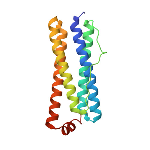
    4XKS, 4XKT, 4XKU

  • PubMed Abstract: 

    Ferritins are iron storage proteins that overcome the problems of toxicity and poor bioavailability of iron by catalyzing iron oxidation and mineralization through the activity of a diiron ferroxidase site. Unlike in other ferritins, the oxidized di-Fe(3+) site of Escherichia coli bacterioferritin (EcBFR) is stable and therefore does not function as a conduit for the transfer of Fe(3+) into the storage cavity, but instead acts as a true catalytic cofactor that cycles its oxidation state while driving Fe(2+) oxidation in the cavity. Herein, we demonstrate that EcBFR mineralization depends on three aromatic residues near the diiron site, Tyr25, Tyr58, and Trp133, and that a transient radical is formed on Tyr25. The data indicate that the aromatic residues, together with a previously identified inner surface iron site, promote mineralization by ensuring the simultaneous delivery of two electrons, derived from Fe(2+) oxidation in the BFR cavity, to the di-ferric catalytic site for safe reduction of O2.


  • Organizational Affiliation

    Centre for Molecular and Structural Biochemistry, School of Chemistry, University of East Anglia, Norwich Research Park, Norwich, NR4 7TJ (UK).


Macromolecules
Find similar proteins by:  (by identity cutoff)  |  3D Structure
Entity ID: 1
MoleculeChains Sequence LengthOrganismDetailsImage
Bacterioferritin
A, B, C, D, E
A, B, C, D, E, F, G, H, I, J, K, L
158Escherichia coli K-12Mutation(s): 1 
Gene Names: bfrb3336JW3298
EC: 1.16.3.1
UniProt
Find proteins for P0ABD3 (Escherichia coli (strain K12))
Explore P0ABD3 
Go to UniProtKB:  P0ABD3
Entity Groups  
Sequence Clusters30% Identity50% Identity70% Identity90% Identity95% Identity100% Identity
UniProt GroupP0ABD3
Sequence Annotations
Expand
  • Reference Sequence
Small Molecules
Ligands 1 Unique
IDChains Name / Formula / InChI Key2D Diagram3D Interactions
SO4
Query on SO4

Download Ideal Coordinates CCD File 
M [auth A]
N [auth A]
O [auth C]
P [auth C]
Q [auth D]
M [auth A],
N [auth A],
O [auth C],
P [auth C],
Q [auth D],
R [auth D],
S [auth E],
T [auth E],
U [auth F],
V [auth G],
W [auth H],
X [auth K]
SULFATE ION
O4 S
QAOWNCQODCNURD-UHFFFAOYSA-L
Experimental Data & Validation

Experimental Data

  • Method: X-RAY DIFFRACTION
  • Resolution: 1.57 Å
  • R-Value Free: 0.172 
  • R-Value Work: 0.149 
  • R-Value Observed: 0.150 
  • Space Group: C 2 2 21
Unit Cell:
Length ( Å )Angle ( ˚ )
a = 127.27α = 90
b = 196.01β = 90
c = 201.87γ = 90
Software Package:
Software NamePurpose
PHENIXrefinement
xia2data reduction
xia2data scaling
PHENIXphasing

Structure Validation

View Full Validation Report


Entry History & Funding Information

Deposition Data


Funding OrganizationLocationGrant Number
Biotechnology and Biological Sciences Research CouncilUnited KingdomBB/IO21884/1

Revision History  (Full details and data files)

  • Version 1.0: 2015-12-16
    Type: Initial release
  • Version 1.1: 2016-01-13
    Changes: Database references
  • Version 1.2: 2016-01-20
    Changes: Database references
  • Version 2.0: 2017-08-30
    Changes: Advisory, Atomic model, Author supporting evidence, Derived calculations
  • Version 2.1: 2024-01-10
    Changes: Data collection, Database references, Refinement description