4XHQ

Re-refinement the crystal structure of Dscam1 isoform 1.34, N-terminal four Ig domains


Experimental Data Snapshot

  • Method: X-RAY DIFFRACTION
  • Resolution: 1.95 Å
  • R-Value Free: 0.233 
  • R-Value Work: 0.203 
  • R-Value Observed: 0.205 

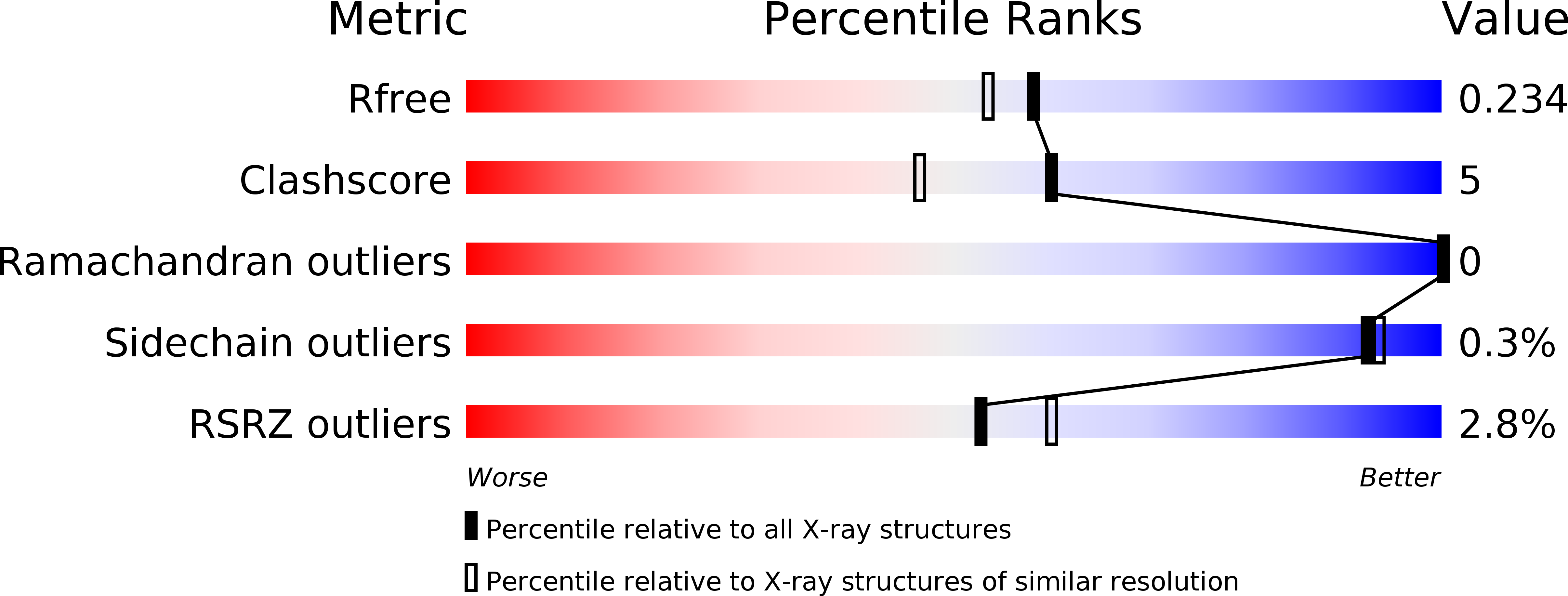
wwPDB Validation   3D Report Full Report


This is version 2.0 of the entry. See complete history

Re-refinement Note

This entry reflects an alternative modeling of the original data in: 2V5M


Literature

Structural basis of Dscam1 homodimerization: Insights into context constraint for protein recognition

Li, S.A.Cheng, L.Yu, Y.Chen, Q.

(2016) Sci Adv 2: e1501118-e1501118

  • DOI: https://doi.org/10.1126/sciadv.1501118
  • Primary Citation of Related Structures:  
    4WVR, 4X5L, 4X83, 4X8X, 4X9B, 4X9F, 4X9G, 4X9H, 4X9I, 4XB7, 4XB8, 4XHQ

  • PubMed Abstract: 

    The Drosophila neural receptor Dscam1 (Down syndrome cell adhesion molecule 1) plays an essential role in neuronal wiring and self-avoidance. Dscam1 potentially encodes 19,008 ectodomains through alternative RNA splicing and exhibits exquisite isoform-specific homophilic binding, which makes it an exceptional example for studying protein binding specificity. However, structural information on Dscam1 is limited, which hinders illumination of the mechanism of Dscam1 isoform-specific recognition. Whether different Dscam1 isoforms adopt the same dimerization mode remains a subject of debate. We present 12 Dscam1 crystal structures, provide direct evidence indicating that all isoforms adopt a conserved homodimer geometry in a modular fashion, identify two mechanisms for the Ig2 binding domain to dispel electrostatic repulsion during dimerization, decode Ig2 binding specificity by a central motif at its symmetry center, uncover the role of glycosylation in Dscam1 homodimerization, and find electrostatic potential complementarity to help define the binding region and the antiparallel binding mode. We then propose a concept that the context of a protein may set restrictions to regulate its binding specificity, which provides a better understanding of protein recognition.


  • Organizational Affiliation

    State Key Laboratory of Biotherapy and Cancer Center, West China Hospital, Sichuan University, and Collaborative Innovation Center of Biotherapy, Chengdu 610041, P. R. China.


Macromolecules
Find similar proteins by:  (by identity cutoff)  |  3D Structure
Entity ID: 1
MoleculeChains Sequence LengthOrganismDetailsImage
Dscam388Drosophila melanogasterMutation(s): 20 
Gene Names: Dscam1DscamCG17800
UniProt
Find proteins for Q0E9H9 (Drosophila melanogaster)
Explore Q0E9H9 
Go to UniProtKB:  Q0E9H9
Entity Groups  
Sequence Clusters30% Identity50% Identity70% Identity90% Identity95% Identity100% Identity
UniProt GroupQ0E9H9
Sequence Annotations
Expand
  • Reference Sequence
Oligosaccharides

Help

Entity ID: 2
MoleculeChains Length2D Diagram Glycosylation3D Interactions
beta-D-mannopyranose-(1-4)-2-acetamido-2-deoxy-beta-D-glucopyranose-(1-4)-2-acetamido-2-deoxy-beta-D-glucopyranose
B
3N-Glycosylation
Glycosylation Resources
GlyTouCan:  G15407YE
GlyCosmos:  G15407YE
GlyGen:  G15407YE
Entity ID: 3
MoleculeChains Length2D Diagram Glycosylation3D Interactions
alpha-D-mannopyranose-(1-3)-[alpha-D-mannopyranose-(1-6)]beta-D-mannopyranose-(1-4)-2-acetamido-2-deoxy-beta-D-glucopyranose-(1-4)-2-acetamido-2-deoxy-beta-D-glucopyranose
C
5N-Glycosylation
Glycosylation Resources
GlyTouCan:  G22768VO
GlyCosmos:  G22768VO
GlyGen:  G22768VO
Small Molecules
Ligands 4 Unique
IDChains Name / Formula / InChI Key2D Diagram3D Interactions
SO4
Query on SO4

Download Ideal Coordinates CCD File 
N [auth A],
O [auth A]
SULFATE ION
O4 S
QAOWNCQODCNURD-UHFFFAOYSA-L
GOL
Query on GOL

Download Ideal Coordinates CCD File 
D [auth A],
E [auth A],
H [auth A]
GLYCEROL
C3 H8 O3
PEDCQBHIVMGVHV-UHFFFAOYSA-N
CL
Query on CL

Download Ideal Coordinates CCD File 
F [auth A],
G [auth A],
I [auth A],
P [auth A]
CHLORIDE ION
Cl
VEXZGXHMUGYJMC-UHFFFAOYSA-M
NA
Query on NA

Download Ideal Coordinates CCD File 
J [auth A],
K [auth A],
L [auth A],
M [auth A]
SODIUM ION
Na
FKNQFGJONOIPTF-UHFFFAOYSA-N
Experimental Data & Validation

Experimental Data

  • Method: X-RAY DIFFRACTION
  • Resolution: 1.95 Å
  • R-Value Free: 0.233 
  • R-Value Work: 0.203 
  • R-Value Observed: 0.205 
  • Space Group: P 42 2 2
Unit Cell:
Length ( Å )Angle ( ˚ )
a = 99.172α = 90
b = 99.172β = 90
c = 163.991γ = 90
Software Package:
Software NamePurpose
PHENIXrefinement
DENZOdata reduction
SCALEPACKdata scaling
PHASERphasing

Structure Validation

View Full Validation Report


Entry History 

Deposition Data

Revision History  (Full details and data files)

  • Version 1.0: 2016-10-12
    Type: Initial release
  • Version 2.0: 2020-07-29
    Type: Remediation
    Reason: Carbohydrate remediation
    Changes: Atomic model, Data collection, Derived calculations, Structure summary