4XCG

Crystal structure of a hexadecameric TF55 complex from S. solfataricus, crystal form I


Experimental Data Snapshot

  • Method: X-RAY DIFFRACTION
  • Resolution: 3.74 Å
  • R-Value Free: 0.292 
  • R-Value Work: 0.247 
  • R-Value Observed: 0.250 

wwPDB Validation   3D Report Full Report


Ligand Structure Quality Assessment 


This is version 1.5 of the entry. See complete history


Literature

Structural and Functional Insights into the Evolution and Stress Adaptation of Type II Chaperonins.

Chaston, J.J.Smits, C.Aragao, D.Wong, A.S.Ahsan, B.Sandin, S.Molugu, S.K.Molugu, S.K.Bernal, R.A.Stock, D.Stewart, A.G.

(2016) Structure 24: 364-374

  • DOI: https://doi.org/10.1016/j.str.2015.12.016
  • Primary Citation of Related Structures:  
    4XCD, 4XCG, 4XCI

  • PubMed Abstract: 

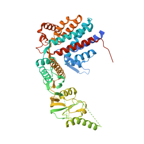
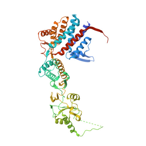
    Chaperonins are essential biological complexes assisting protein folding in all kingdoms of life. Whereas homooligomeric bacterial GroEL binds hydrophobic substrates non-specifically, the heterooligomeric eukaryotic CCT binds specifically to distinct classes of substrates. Sulfolobales, which survive in a wide range of temperatures, have evolved three different chaperonin subunits (α, β, γ) that form three distinct complexes tailored for different substrate classes at cold, normal, and elevated temperatures. The larger octadecameric β complexes cater for substrates under heat stress, whereas smaller hexadecameric αβ complexes prevail under normal conditions. The cold-shock complex contains all three subunits, consistent with greater substrate specificity. Structural analysis using crystallography and electron microscopy reveals the geometry of these complexes and shows a novel arrangement of the α and β subunits in the hexadecamer enabling incorporation of the γ subunit.


  • Organizational Affiliation

    Molecular, Structural and Computational Biology Division, The Victor Chang Cardiac Research Institute, Darlinghurst, NSW 2010, Australia; Faculty of Medicine, The University of New South Wales, Sydney, NSW 2052, Australia.


Macromolecules
Find similar proteins by:  (by identity cutoff)  |  3D Structure
Entity ID: 1
MoleculeChains Sequence LengthOrganismDetailsImage
Thermosome subunit betaA [auth B]557Saccharolobus solfataricus P2Mutation(s): 0 
UniProt
Find proteins for Q9V2T8 (Saccharolobus solfataricus (strain ATCC 35092 / DSM 1617 / JCM 11322 / P2))
Explore Q9V2T8 
Go to UniProtKB:  Q9V2T8
Entity Groups  
Sequence Clusters30% Identity50% Identity70% Identity90% Identity95% Identity100% Identity
UniProt GroupQ9V2T8
Sequence Annotations
Expand
  • Reference Sequence
Find similar proteins by:  (by identity cutoff)  |  3D Structure
Entity ID: 2
MoleculeChains Sequence LengthOrganismDetailsImage
Thermosome subunit alphaB [auth A]559Saccharolobus solfataricus P2Mutation(s): 0 
UniProt
Find proteins for Q9V2S9 (Saccharolobus solfataricus (strain ATCC 35092 / DSM 1617 / JCM 11322 / P2))
Explore Q9V2S9 
Go to UniProtKB:  Q9V2S9
Entity Groups  
Sequence Clusters30% Identity50% Identity70% Identity90% Identity95% Identity100% Identity
UniProt GroupQ9V2S9
Sequence Annotations
Expand
  • Reference Sequence
Small Molecules
Ligands 1 Unique
IDChains Name / Formula / InChI Key2D Diagram3D Interactions
ADP
Query on ADP

Download Ideal Coordinates CCD File 
C [auth B]ADENOSINE-5'-DIPHOSPHATE
C10 H15 N5 O10 P2
XTWYTFMLZFPYCI-KQYNXXCUSA-N
Experimental Data & Validation

Experimental Data

  • Method: X-RAY DIFFRACTION
  • Resolution: 3.74 Å
  • R-Value Free: 0.292 
  • R-Value Work: 0.247 
  • R-Value Observed: 0.250 
  • Space Group: I 4 2 2
Unit Cell:
Length ( Å )Angle ( ˚ )
a = 145.776α = 90
b = 145.776β = 90
c = 248.092γ = 90
Software Package:
Software NamePurpose
PHENIXrefinement
PDB_EXTRACTdata extraction
PHASERphasing
Aimlessdata scaling

Structure Validation

View Full Validation Report


Ligand Structure Quality Assessment 


Entry History & Funding Information

Deposition Data


Funding OrganizationLocationGrant Number
Australian Research Council (ARC)AustraliaDP1093649

Revision History  (Full details and data files)

  • Version 1.0: 2016-02-10
    Type: Initial release
  • Version 1.1: 2016-02-24
    Changes: Database references
  • Version 1.2: 2016-03-09
    Changes: Database references
  • Version 1.3: 2017-11-01
    Changes: Author supporting evidence, Data collection, Database references, Derived calculations, Refinement description
  • Version 1.4: 2020-01-01
    Changes: Author supporting evidence
  • Version 1.5: 2023-09-27
    Changes: Data collection, Database references, Refinement description