4X97

Crystal structure of Lysosomal Phospholipase A2 in complex with methyl arachidonyl fluorophosphonate (MAFP)

  • Classification: TRANSFERASE
  • Organism(s): Homo sapiens
  • Expression System: Homo sapiens
  • Mutation(s): No 

  • Deposited: 2014-12-11 Released: 2015-03-11 
  • Deposition Author(s): Glukhova, A., Tesmer, J.J.G.
  • Funding Organization(s): National Institutes of Health/National Heart, Lung, and Blood Institute (NIH/NHLBI)

Experimental Data Snapshot

  • Method: X-RAY DIFFRACTION
  • Resolution: 2.65 Å
  • R-Value Free: 0.219 
  • R-Value Work: 0.179 
  • R-Value Observed: 0.181 

wwPDB Validation   3D Report Full Report


Ligand Structure Quality Assessment 


This is version 1.6 of the entry. See complete history


Literature

Structure and function of lysosomal phospholipase A2 and lecithin:cholesterol acyltransferase.

Glukhova, A.Hinkovska-Galcheva, V.Kelly, R.Abe, A.Shayman, J.A.Tesmer, J.J.

(2015) Nat Commun 6: 6250-6250

  • DOI: https://doi.org/10.1038/ncomms7250
  • Primary Citation of Related Structures:  
    4X90, 4X91, 4X92, 4X93, 4X94, 4X95, 4X96, 4X97

  • PubMed Abstract: 

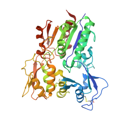
    Lysosomal phospholipase A2 (LPLA2) and lecithin:cholesterol acyltransferase (LCAT) belong to a structurally uncharacterized family of key lipid-metabolizing enzymes responsible for lung surfactant catabolism and for reverse cholesterol transport, respectively. Whereas LPLA2 is predicted to underlie the development of drug-induced phospholipidosis, somatic mutations in LCAT cause fish eye disease and familial LCAT deficiency. Here we describe several high-resolution crystal structures of human LPLA2 and a low-resolution structure of LCAT that confirms its close structural relationship to LPLA2. Insertions in the α/β hydrolase core of LPLA2 form domains that are responsible for membrane interaction and binding the acyl chains and head groups of phospholipid substrates. The LCAT structure suggests the molecular basis underlying human disease for most of the known LCAT missense mutations, and paves the way for rational development of new therapeutics to treat LCAT deficiency, atherosclerosis and acute coronary syndrome.


  • Organizational Affiliation

    1] Life Sciences Institute and the Departments of Pharmacology and Biological Chemistry, University of Michigan, Ann Arbor, Michigan 48109, USA [2] Program in Chemical Biology, University of Michigan, Ann Arbor, Michigan 48109, USA.


Macromolecules
Find similar proteins by:  (by identity cutoff)  |  3D Structure
Entity ID: 1
MoleculeChains Sequence LengthOrganismDetailsImage
Group XV phospholipase A2
A, B, C, D
380Homo sapiensMutation(s): 0 
Gene Names: PLA2G15LYPLA3UNQ341/PRO540
EC: 2.3.1
UniProt & NIH Common Fund Data Resources
Find proteins for Q8NCC3 (Homo sapiens)
Explore Q8NCC3 
Go to UniProtKB:  Q8NCC3
PHAROS:  Q8NCC3
GTEx:  ENSG00000103066 
Entity Groups  
Sequence Clusters30% Identity50% Identity70% Identity90% Identity95% Identity100% Identity
UniProt GroupQ8NCC3
Sequence Annotations
Expand
  • Reference Sequence
Small Molecules
Ligands 5 Unique
IDChains Name / Formula / InChI Key2D Diagram3D Interactions
MAY
Query on MAY

Download Ideal Coordinates CCD File 
EA [auth D],
I [auth A],
P [auth B],
X [auth C]
METHYL ARACHIDONYL FLUOROPHOSPHONATE
C21 H36 F O2 P
KWKZCGMJGHHOKJ-WTIHWRCNSA-N
EPE
Query on EPE

Download Ideal Coordinates CCD File 
FA [auth D],
J [auth A],
Q [auth B],
Y [auth C]
4-(2-HYDROXYETHYL)-1-PIPERAZINE ETHANESULFONIC ACID
C8 H18 N2 O4 S
JKMHFZQWWAIEOD-UHFFFAOYSA-N
NAG
Query on NAG

Download Ideal Coordinates CCD File 
AA [auth D]
BA [auth D]
CA [auth D]
DA [auth D]
E [auth A]
AA [auth D],
BA [auth D],
CA [auth D],
DA [auth D],
E [auth A],
F [auth A],
G [auth A],
H [auth A],
L [auth B],
M [auth B],
N [auth B],
O [auth B],
T [auth C],
U [auth C],
V [auth C],
W [auth C]
2-acetamido-2-deoxy-beta-D-glucopyranose
C8 H15 N O6
OVRNDRQMDRJTHS-FMDGEEDCSA-N
PO4
Query on PO4

Download Ideal Coordinates CCD File 
GA [auth D],
K [auth A],
S [auth B],
Z [auth C]
PHOSPHATE ION
O4 P
NBIIXXVUZAFLBC-UHFFFAOYSA-K
CL
Query on CL

Download Ideal Coordinates CCD File 
R [auth B]CHLORIDE ION
Cl
VEXZGXHMUGYJMC-UHFFFAOYSA-M
Experimental Data & Validation

Experimental Data

  • Method: X-RAY DIFFRACTION
  • Resolution: 2.65 Å
  • R-Value Free: 0.219 
  • R-Value Work: 0.179 
  • R-Value Observed: 0.181 
  • Space Group: P 1
Unit Cell:
Length ( Å )Angle ( ˚ )
a = 69.147α = 88.85
b = 85.495β = 70.87
c = 88.852γ = 79.74
Software Package:
Software NamePurpose
HKL-2000data scaling
SCALEPACKdata scaling
Aimlessdata scaling
Cootmodel building
PHASERphasing
REFMACrefinement
PDB_EXTRACTdata extraction

Structure Validation

View Full Validation Report


Ligand Structure Quality Assessment 


Entry History & Funding Information

Deposition Data


Funding OrganizationLocationGrant Number
National Institutes of Health/National Heart, Lung, and Blood Institute (NIH/NHLBI)United StatesHL086865

Revision History  (Full details and data files)

  • Version 1.0: 2015-03-11
    Type: Initial release
  • Version 1.1: 2015-03-18
    Changes: Database references
  • Version 1.2: 2017-09-13
    Changes: Advisory, Author supporting evidence, Derived calculations, Source and taxonomy
  • Version 1.3: 2017-11-22
    Changes: Refinement description
  • Version 1.4: 2019-12-04
    Changes: Author supporting evidence
  • Version 1.5: 2020-07-29
    Type: Remediation
    Reason: Carbohydrate remediation
    Changes: Data collection, Derived calculations, Structure summary
  • Version 1.6: 2023-09-27
    Changes: Data collection, Database references, Refinement description, Structure summary