4WHO

Resting Protocatechuate 3,4-dioxygenase (pseudomonas putida) at pH 8.5


Experimental Data Snapshot

  • Method: X-RAY DIFFRACTION
  • Resolution: 1.83 Å
  • R-Value Free: 0.216 
  • R-Value Work: 0.180 
  • R-Value Observed: 0.182 

wwPDB Validation   3D Report Full Report


This is version 1.6 of the entry. See complete history


Literature

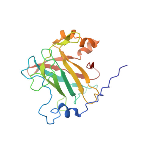
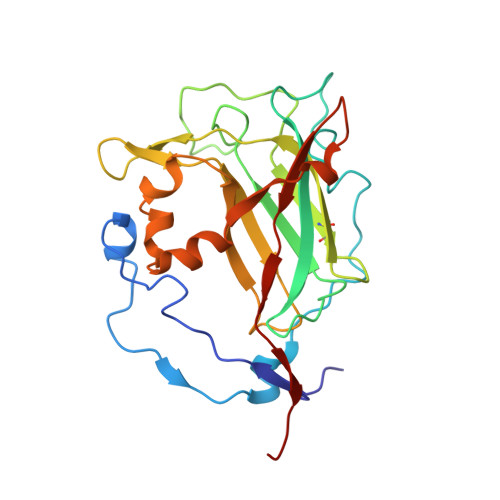
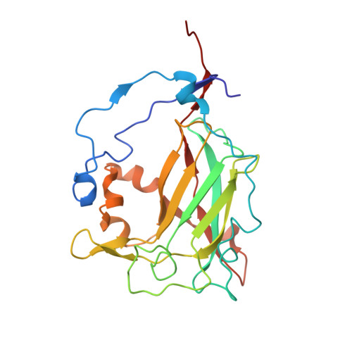
Crystal structures of alkylperoxo and anhydride intermediates in an intradiol ring-cleaving dioxygenase.

Knoot, C.J.Purpero, V.M.Lipscomb, J.D.

(2015) Proc Natl Acad Sci U S A 112: 388-393

  • DOI: https://doi.org/10.1073/pnas.1419118112
  • Primary Citation of Related Structures:  
    4WHO, 4WHP, 4WHQ, 4WHR, 4WHS

  • PubMed Abstract: 

    Intradiol aromatic ring-cleaving dioxygenases use an active site, nonheme Fe(3+) to activate O2 and catecholic substrates for reaction. The inability of Fe(3+) to directly bind O2 presents a mechanistic conundrum. The reaction mechanism of protocatechuate 3,4-dioxygenase is investigated here using the alternative substrate 4-fluorocatechol. This substrate is found to slow the reaction at several steps throughout the mechanistic cycle, allowing the intermediates to be detected in solution studies. When the reaction was initiated in an enzyme crystal, it was found to halt at one of two intermediates depending on the pH of the surrounding solution. The X-ray crystal structure of the intermediate at pH 6.5 revealed the key alkylperoxo-Fe(3+) species, and the anhydride-Fe(3+) intermediate was found for a crystal reacted at pH 8.5. Intermediates of these types have not been structurally characterized for intradiol dioxygenases, and they validate four decades of spectroscopic, kinetic, and computational studies. In contrast to our similar in crystallo crystallographic studies of an Fe(2+)-containing extradiol dioxygenase, no evidence for a superoxo or peroxo intermediate preceding the alkylperoxo was found. This observation and the lack of spectroscopic evidence for an Fe(2+) intermediate that could bind O2 are consistent with concerted formation of the alkylperoxo followed by Criegee rearrangement to yield the anhydride and ultimately ring-opened product. Structural comparison of the alkylperoxo intermediates from the intra- and extradiol dioxygenases provides a rationale for site specificity of ring cleavage.


  • Organizational Affiliation

    Department of Biochemistry Molecular Biology and Biophysics and Center for Metals in Biocatalysis, University of Minnesota, Minneapolis, MN 55455.


Macromolecules
Find similar proteins by:  (by identity cutoff)  |  3D Structure
Entity ID: 1
MoleculeChains Sequence LengthOrganismDetailsImage
Protocatechuate 3,4-dioxygenase alpha chainA,
B [auth E],
C
200Pseudomonas putidaMutation(s): 0 
Gene Names: pcaG
EC: 1.13.11.3
UniProt
Find proteins for P00436 (Pseudomonas putida)
Explore P00436 
Go to UniProtKB:  P00436
Entity Groups  
Sequence Clusters30% Identity50% Identity70% Identity90% Identity95% Identity100% Identity
UniProt GroupP00436
Sequence Annotations
Expand
  • Reference Sequence
Find similar proteins by:  (by identity cutoff)  |  3D Structure
Entity ID: 2
MoleculeChains Sequence LengthOrganismDetailsImage
Protocatechuate 3,4-dioxygenase beta chainD [auth B]238Pseudomonas putidaMutation(s): 0 
Gene Names: pcaH
EC: 1.13.11.3
UniProt
Find proteins for P00437 (Pseudomonas putida)
Explore P00437 
Go to UniProtKB:  P00437
Entity Groups  
Sequence Clusters30% Identity50% Identity70% Identity90% Identity95% Identity100% Identity
UniProt GroupP00437
Sequence Annotations
Expand
  • Reference Sequence
Find similar proteins by:  (by identity cutoff)  |  3D Structure
Entity ID: 3
MoleculeChains Sequence LengthOrganismDetailsImage
Protocatechuate 3,4-dioxygenase beta chainE [auth D],
F
238Pseudomonas putidaMutation(s): 0 
Gene Names: pcaH
EC: 1.13.11.3
UniProt
Find proteins for P00437 (Pseudomonas putida)
Explore P00437 
Go to UniProtKB:  P00437
Entity Groups  
Sequence Clusters30% Identity50% Identity70% Identity90% Identity95% Identity100% Identity
UniProt GroupP00437
Sequence Annotations
Expand
  • Reference Sequence
Experimental Data & Validation

Experimental Data

  • Method: X-RAY DIFFRACTION
  • Resolution: 1.83 Å
  • R-Value Free: 0.216 
  • R-Value Work: 0.180 
  • R-Value Observed: 0.182 
  • Space Group: I 2 2 2
Unit Cell:
Length ( Å )Angle ( ˚ )
a = 128.092α = 90
b = 140.636β = 90
c = 168.66γ = 90
Software Package:
Software NamePurpose
HKL-3000data reduction
REFMACrefinement
PDB_EXTRACTdata extraction
HKL-3000data scaling
DENZOdata reduction
SCALEPACKdata scaling

Structure Validation

View Full Validation Report


Entry History & Funding Information

Deposition Data


Funding OrganizationLocationGrant Number
National Institutes of Health/National Institute of General Medical Sciences (NIH/NIGMS)United StatesGM24689
National Institutes of Health/National Institute of General Medical Sciences (NIH/NIGMS)United StatesGM08700

Revision History  (Full details and data files)

  • Version 1.0: 2014-12-31
    Type: Initial release
  • Version 1.1: 2015-01-14
    Changes: Database references
  • Version 1.2: 2015-01-28
    Changes: Database references
  • Version 1.3: 2017-09-27
    Changes: Author supporting evidence, Database references, Derived calculations, Other, Refinement description, Source and taxonomy
  • Version 1.4: 2019-12-25
    Changes: Author supporting evidence
  • Version 1.5: 2023-09-27
    Changes: Data collection, Database references, Derived calculations, Refinement description
  • Version 1.6: 2023-11-15
    Changes: Data collection