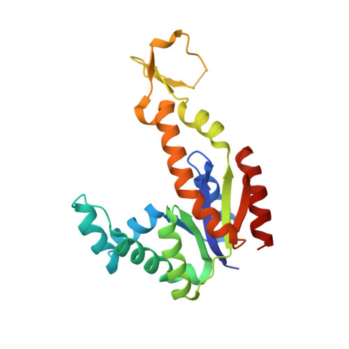
4W5H

New structural conformations of adenylate kinase from Streptococcus pneumoniae D39


Experimental Data Snapshot

  • Method: X-RAY DIFFRACTION
  • Resolution: 1.96 Å
  • R-Value Free: 0.219 
  • R-Value Work: 0.177 
  • R-Value Observed: 0.181 

wwPDB Validation   3D Report Full Report


This is version 1.2 of the entry. See complete history


Literature

New crystal structures of adenylate kinase from Streptococcus pneumoniae D39 in two conformations.

Thach, T.T.Lee, S.

(2014) Acta Crystallogr F Struct Biol Commun 70: 1468-1471

  • DOI: https://doi.org/10.1107/S2053230X14020718
  • Primary Citation of Related Structures:  
    4W5H, 4W5J

  • PubMed Abstract: 

    Adenylate kinases (AdKs; EC 2.7.3.4) play a critical role in intercellular homeostasis by the interconversion of ATP and AMP to two ADP molecules. Crystal structures of adenylate kinase from Streptococcus pneumoniae D39 (SpAdK) have recently been determined using ligand-free and inhibitor-bound crystals belonging to space groups P21 and P1, respectively. Here, new crystal structures of SpAdK in ligand-free and inhibitor-bound states determined at 1.96 and 1.65 Å resolution, respectively, are reported. The new ligand-free crystal belonged to space group C2, with unit-cell parameters a=73.5, b=54.3, c=62.7 Å, β=118.8°. The new ligand-free structure revealed an open conformation that differed from the previously determined conformation, with an r.m.s.d on Cα atoms of 1.4 Å. The new crystal of the complex with the two-substrate-mimicking inhibitor P1,P5-bis(adenosine-5'-)pentaphosphate (Ap5A) belonged to space group P1, with unit-cell parameters a=53.9, b=62.3, c=63.0 Å, α=101.9, β=112.6, γ=89.9°. Despite belonging to the same space group as the previously reported crystal, the new Ap5A-bound crystal contains four molecules in the asymmetric unit, compared with two in the previous crystal, and shows slightly different lattice contacts. These results demonstrate that SpAdK can crystallize promiscuously in different forms and that the open structure is flexible in conformation.


  • Organizational Affiliation

    Department of Biological Sciences, Sungkyunkwan University, 2066 Seobu-ro, Suwon, Gyeonggi 440-746, Republic of Korea.


Macromolecules
Find similar proteins by:  (by identity cutoff)  |  3D Structure
Entity ID: 1
MoleculeChains Sequence LengthOrganismDetailsImage
Adenylate kinase217Streptococcus pneumoniae D39Mutation(s): 0 
Gene Names: adkSPD_0214
EC: 2.7.4.3
UniProt
Find proteins for Q04ML5 (Streptococcus pneumoniae serotype 2 (strain D39 / NCTC 7466))
Explore Q04ML5 
Go to UniProtKB:  Q04ML5
Entity Groups  
Sequence Clusters30% Identity50% Identity70% Identity90% Identity95% Identity100% Identity
UniProt GroupQ04ML5
Sequence Annotations
Expand
  • Reference Sequence
Small Molecules
Ligands 2 Unique
IDChains Name / Formula / InChI Key2D Diagram3D Interactions
EPE
Query on EPE

Download Ideal Coordinates CCD File 
C [auth A]4-(2-HYDROXYETHYL)-1-PIPERAZINE ETHANESULFONIC ACID
C8 H18 N2 O4 S
JKMHFZQWWAIEOD-UHFFFAOYSA-N
TRS
Query on TRS

Download Ideal Coordinates CCD File 
B [auth A],
D [auth A]
2-AMINO-2-HYDROXYMETHYL-PROPANE-1,3-DIOL
C4 H12 N O3
LENZDBCJOHFCAS-UHFFFAOYSA-O
Experimental Data & Validation

Experimental Data

  • Method: X-RAY DIFFRACTION
  • Resolution: 1.96 Å
  • R-Value Free: 0.219 
  • R-Value Work: 0.177 
  • R-Value Observed: 0.181 
  • Space Group: C 1 2 1
Unit Cell:
Length ( Å )Angle ( ˚ )
a = 73.508α = 90
b = 54.331β = 118.85
c = 62.711γ = 90
Software Package:
Software NamePurpose
PHENIXrefinement
PDB_EXTRACTdata extraction

Structure Validation

View Full Validation Report


Entry History 

Deposition Data

Revision History  (Full details and data files)

  • Version 1.0: 2014-11-26
    Type: Initial release
  • Version 1.1: 2017-08-16
    Changes: Database references, Derived calculations, Other, Source and taxonomy
  • Version 1.2: 2023-11-08
    Changes: Data collection, Database references, Refinement description