Structures of Two Bacterial Resistance Factors Mediating tRNA-Dependent Aminoacylation of Phosphatidylglycerol with Lysine or Alanine.
Hebecker, S., Krausze, J., Hasenkampf, T., Schneider, J., Groenewold, M., Reichelt, J., Jahn, D., Heinz, D.W., Moser, J.(2015) Proc Natl Acad Sci U S A 112: 10691
- PubMed: 26261323
- DOI: https://doi.org/10.1073/pnas.1511167112
- Primary Citation of Related Structures:
4V34, 4V35, 4V36 - PubMed Abstract:
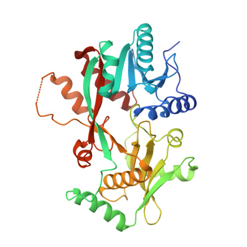
The cytoplasmic membrane is probably the most important physical barrier between microbes and the surrounding habitat. Aminoacylation of the polar head group of the phospholipid phosphatidylglycerol (PG) catalyzed by Ala-tRNA(Ala)-dependent alanyl-phosphatidylglycerol synthase (A-PGS) or by Lys-tRNA(Lys)-dependent lysyl-phosphatidylglycerol synthase (L-PGS) enables bacteria to cope with cationic peptides that are harmful to the integrity of the cell membrane. Accordingly, these synthases also have been designated as multiple peptide resistance factors (MprF). They consist of a separable C-terminal catalytic domain and an N-terminal transmembrane flippase domain. Here we present the X-ray crystallographic structure of the catalytic domain of A-PGS from the opportunistic human pathogen Pseudomonas aeruginosa. In parallel, the structure of the related lysyl-phosphatidylglycerol-specific L-PGS domain from Bacillus licheniformis in complex with the substrate analog L-lysine amide is presented. Both proteins reveal a continuous tunnel that allows the hydrophobic lipid substrate PG and the polar aminoacyl-tRNA substrate to access the catalytic site from opposite directions. Substrate recognition of A-PGS versus L-PGS was investigated using misacylated tRNA variants. The structural work presented here in combination with biochemical experiments using artificial tRNA or artificial lipid substrates reveals the tRNA acceptor stem, the aminoacyl moiety, and the polar head group of PG as the main determinants for substrate recognition. A mutagenesis approach yielded the complementary amino acid determinants of tRNA interaction. These results have broad implications for the design of L-PGS and A-PGS inhibitors that could render microbial pathogens more susceptible to antimicrobial compounds.
Organizational Affiliation:
Institute of Microbiology, Technische Universität Braunschweig, 38106 Braunschweig, Germany;