Role of Local Structure and Dynamics of Small Ligand Migration in Proteins: A Study of a Mutated Truncated Hemoprotein from Thermobifida Fusca by Time Resolved Mir Spectroscopy.
Patrizi, B., Lapini, A., Di Donato, M., Marcelli, A., Lima, M., Righini, R., Foggi, P., Baiocco, P., Bonamore, A., Boffi, A.(2014) J Phys Chem B 118: 9209
- PubMed: 25019316
- DOI: https://doi.org/10.1021/jp504499b
- Primary Citation of Related Structures:
4UZV - PubMed Abstract:
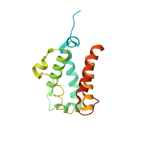
Carbon monoxide recombination dynamics in a mutant of the truncated hemoglobin from Thermobida fusca (3F-Tf-trHb) has been analyzed by means of ultrafast Visible-pump/MidIR-probe spectroscopy and compared with that of the wild-type protein. In 3F-Tf-trHb, three topologically relevant amino acids, responsible for the ligand stabilization through the formation of a H-bond network (TyrB10 TyrCD1 and TrpG8), have been replaced by Phe residues. X-ray diffraction data show that Phe residues in positions B10 and G8 maintain the same rotameric arrangements as Tyr and Trp in the wild-type protein, while Phe in position CD1 displays significant rotameric heterogeneity. Photodissociation of the ligand has been induced by exciting the sample with 550 nm pump pulses and the CO rebinding has been monitored in two mid-IR regions respectively corresponding to the ν(CO) stretching vibration of the iron-bound CO (1880-1980 cm(-1)) and of the dissociated free CO (2050-2200 cm(-1)). In both the mutant and wild-type protein, a significant amount of geminate CO rebinding is observed on a subnanosecond time scale. Despite the absence of the distal pocket hydrogen-bonding network, the kinetics of geminate rebinding in 3F-Tf-trHb is very similar to the wild-type, showing how the reactivity of dissociated CO toward the heme is primarily regulated by the effective volume and flexibility of the distal pocket and by caging effects exerted on the free CO on the analyzed time scale.
Organizational Affiliation:
LENS (European Laboratory for Nonlinear Spectroscopy) Via N. Carrara 1, Sesto Fiorentino, Florence 50019, Italy.