4UVM

In meso crystal structure of the POT family transporter PepTSo


Experimental Data Snapshot

  • Method: X-RAY DIFFRACTION
  • Resolution: 3.00 Å
  • R-Value Free: 0.258 
  • R-Value Work: 0.220 
  • R-Value Observed: 0.222 

wwPDB Validation   3D Report Full Report


Ligand Structure Quality Assessment 


This is version 1.3 of the entry. See complete history


Literature

Gating Topology of the Proton-Coupled Oligopeptide Symporters.

Fowler, P.W.Orwick-Rydmark, M.Radestock, S.Solcan, N.Dijkman, P.M.Lyons, J.A.Kwok, J.Caffrey, M.Watts, A.Forrest, L.R.Newstead, S.

(2015) Structure 23: 290

  • DOI: https://doi.org/10.1016/j.str.2014.12.012
  • Primary Citation of Related Structures:  
    4UVM

  • PubMed Abstract: 

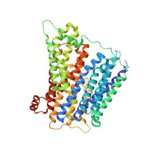
    Proton-coupled oligopeptide transporters belong to the major facilitator superfamily (MFS) of membrane transporters. Recent crystal structures suggest the MFS fold facilitates transport through rearrangement of their two six-helix bundles around a central ligand binding site; how this is achieved, however, is poorly understood. Using modeling, molecular dynamics, crystallography, functional assays, and site-directed spin labeling combined with double electron-electron resonance (DEER) spectroscopy, we present a detailed study of the transport dynamics of two bacterial oligopeptide transporters, PepTSo and PepTSt. Our results identify several salt bridges that stabilize outward-facing conformations and we show that, for all the current structures of MFS transporters, the first two helices of each of the four inverted-topology repeat units form half of either the periplasmic or cytoplasmic gate and that these function cooperatively in a scissor-like motion to control access to the peptide binding site during transport.


  • Organizational Affiliation

    Department of Biochemistry, University of Oxford, South Parks Road, Oxford OX1 3QU, UK. Electronic address: philip.fowler@bioch.ox.ac.uk.


Macromolecules
Find similar proteins by:  (by identity cutoff)  |  3D Structure
Entity ID: 1
MoleculeChains Sequence LengthOrganismDetailsImage
GLUTATHIONE UPTAKE TRANSPORTER524Shewanella oneidensis MR-1Mutation(s): 0 
Membrane Entity: Yes 
UniProt
Find proteins for Q8EKT7 (Shewanella oneidensis (strain MR-1))
Explore Q8EKT7 
Go to UniProtKB:  Q8EKT7
Entity Groups  
Sequence Clusters30% Identity50% Identity70% Identity90% Identity95% Identity100% Identity
UniProt GroupQ8EKT7
Sequence Annotations
Expand
  • Reference Sequence
Experimental Data & Validation

Experimental Data

  • Method: X-RAY DIFFRACTION
  • Resolution: 3.00 Å
  • R-Value Free: 0.258 
  • R-Value Work: 0.220 
  • R-Value Observed: 0.222 
  • Space Group: P 41 21 2
Unit Cell:
Length ( Å )Angle ( ˚ )
a = 86.83α = 90
b = 86.83β = 90
c = 219.82γ = 90
Software Package:
Software NamePurpose
PHENIXrefinement
XDSdata reduction
XSCALEdata scaling
PHASERphasing

Structure Validation

View Full Validation Report


Ligand Structure Quality Assessment 


Entry History 

Deposition Data

Revision History  (Full details and data files)

  • Version 1.0: 2015-02-04
    Type: Initial release
  • Version 1.1: 2015-02-18
    Changes: Database references
  • Version 1.2: 2019-09-25
    Changes: Data collection, Experimental preparation, Other
  • Version 1.3: 2024-01-10
    Changes: Data collection, Database references, Derived calculations, Refinement description