4UBF

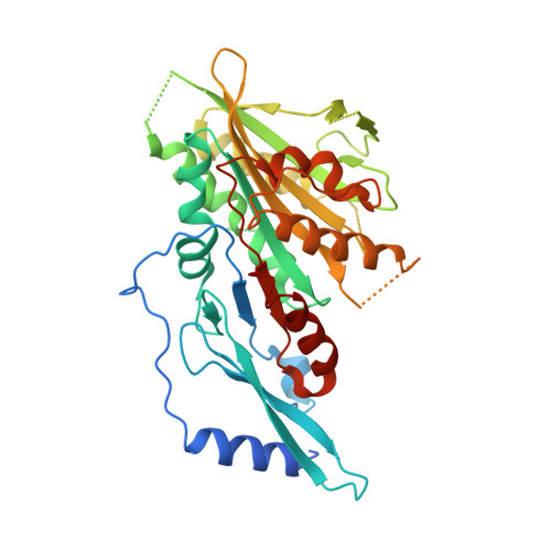
HsMCAK motor domain complex


Experimental Data Snapshot

  • Method: X-RAY DIFFRACTION
  • Resolution: 3.00 Å
  • R-Value Free: 0.286 
  • R-Value Work: 0.264 
  • R-Value Observed: 0.265 

wwPDB Validation   3D Report Full Report


Ligand Structure Quality Assessment 


This is version 1.3 of the entry. See complete history


Literature

The C-terminal region of the motor protein MCAK controls its structure and activity through a conformational switch.

Talapatra, S.K.Harker, B.Welburn, J.P.

(2015) Elife 4

  • DOI: https://doi.org/10.7554/eLife.06421
  • Primary Citation of Related Structures:  
    4UBF

  • PubMed Abstract: 

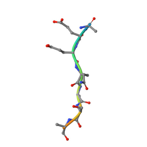
    The precise regulation of microtubule dynamics is essential during cell division. The kinesin-13 motor protein MCAK is a potent microtubule depolymerase. The divergent non-motor regions flanking the ATPase domain are critical in regulating its targeting and activity. However, the molecular basis for the function of the non-motor regions within the context of full-length MCAK is unknown. Here, we determine the structure of MCAK motor domain bound to its regulatory C-terminus. Our analysis reveals that the MCAK C-terminus binds to two motor domains in solution and is displaced allosterically upon microtubule binding, which allows its robust accumulation at microtubule ends. These results demonstrate that MCAK undergoes long-range conformational changes involving its C-terminus during the soluble to microtubule-bound transition and that the C-terminus-motor interaction represents a structural intermediate in the MCAK catalytic cycle. Together, our work reveals intrinsic molecular mechanisms underlying the regulation of kinesin-13 activity.


  • Organizational Affiliation

    Wellcome Trust Centre for Cell Biology, School of Biological Sciences, University of Edinburgh, Edinburgh, United Kingdom.


Macromolecules
Find similar proteins by:  (by identity cutoff)  |  3D Structure
Entity ID: 1
MoleculeChains Sequence LengthOrganismDetailsImage
Kinesin-like protein KIF2C
A, B, C, D
387Homo sapiensMutation(s): 0 
Gene Names: KIF2CKNSL6
UniProt & NIH Common Fund Data Resources
Find proteins for Q99661 (Homo sapiens)
Explore Q99661 
Go to UniProtKB:  Q99661
PHAROS:  Q99661
GTEx:  ENSG00000142945 
Entity Groups  
Sequence Clusters30% Identity50% Identity70% Identity90% Identity95% Identity100% Identity
UniProt GroupQ99661
Sequence Annotations
Expand
  • Reference Sequence

Find similar proteins by:  Sequence   |   3D Structure  

Entity ID: 2
MoleculeChains Sequence LengthOrganismDetailsImage
Kinesin-like protein KIF2CE [auth P]12Homo sapiensMutation(s): 0 
Gene Names: KIF2CKNSL6
UniProt & NIH Common Fund Data Resources
Find proteins for Q99661 (Homo sapiens)
Explore Q99661 
Go to UniProtKB:  Q99661
PHAROS:  Q99661
GTEx:  ENSG00000142945 
Entity Groups  
Sequence Clusters30% Identity50% Identity70% Identity90% Identity95% Identity100% Identity
UniProt GroupQ99661
Sequence Annotations
Expand
  • Reference Sequence
Small Molecules
Ligands 2 Unique
IDChains Name / Formula / InChI Key2D Diagram3D Interactions
ADP
Query on ADP

Download Ideal Coordinates CCD File 
G [auth A],
I [auth B],
M [auth C],
P [auth D]
ADENOSINE-5'-DIPHOSPHATE
C10 H15 N5 O10 P2
XTWYTFMLZFPYCI-KQYNXXCUSA-N
MG
Query on MG

Download Ideal Coordinates CCD File 
F [auth A]
H [auth A]
J [auth B]
K [auth B]
L [auth C]
F [auth A],
H [auth A],
J [auth B],
K [auth B],
L [auth C],
N [auth C],
O [auth D]
MAGNESIUM ION
Mg
JLVVSXFLKOJNIY-UHFFFAOYSA-N
Experimental Data & Validation

Experimental Data

  • Method: X-RAY DIFFRACTION
  • Resolution: 3.00 Å
  • R-Value Free: 0.286 
  • R-Value Work: 0.264 
  • R-Value Observed: 0.265 
  • Space Group: P 1 21 1
Unit Cell:
Length ( Å )Angle ( ˚ )
a = 46.306α = 90
b = 245.644β = 95.84
c = 79.399γ = 90
Software Package:
Software NamePurpose
PHENIXrefinement

Structure Validation

View Full Validation Report


Ligand Structure Quality Assessment 


Entry History & Funding Information

Deposition Data


Funding OrganizationLocationGrant Number
Cancer Research UKUnited Kingdom543WJW_R42094

Revision History  (Full details and data files)

  • Version 1.0: 2015-05-06
    Type: Initial release
  • Version 1.1: 2017-08-30
    Changes: Author supporting evidence
  • Version 1.2: 2018-06-06
    Changes: Data collection, Database references
  • Version 1.3: 2023-12-20
    Changes: Data collection, Database references, Derived calculations, Refinement description