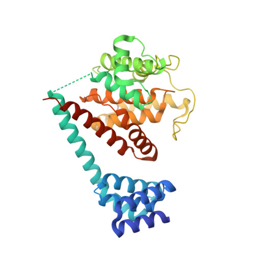
4U04

Structure of a eukaryotic fic domain containing protein


Experimental Data Snapshot

  • Method: X-RAY DIFFRACTION
  • Resolution: 2.48 Å
  • R-Value Free: 0.245 
  • R-Value Work: 0.218 
  • R-Value Observed: 0.219 

wwPDB Validation   3D Report Full Report


This is version 1.2 of the entry. See complete history


Literature

Crystal structure of the human, FIC-domain containing protein HYPE and implications for its functions.

Bunney, T.D.Cole, A.R.Broncel, M.Esposito, D.Tate, E.W.Katan, M.

(2014) Structure 22: 1831-1843

  • DOI: https://doi.org/10.1016/j.str.2014.10.007
  • Primary Citation of Related Structures:  
    4U04, 4U07, 4U0S, 4U0U, 4U0Z

  • PubMed Abstract: 

    Protein AMPylation, the transfer of AMP from ATP to protein targets, has been recognized as a new mechanism of host-cell disruption by some bacterial effectors that typically contain a FIC-domain. Eukaryotic genomes also encode one FIC-domain protein,HYPE, which has remained poorly characterized.Here we describe the structure of human HYPE, solved by X-ray crystallography, representing the first structure of a eukaryotic FIC-domain protein. We demonstrate that HYPE forms stable dimers with structurally and functionally integrated FIC-domains and with TPR-motifs exposed for protein-protein interactions. As HYPE also uniquely possesses a transmembrane helix, dimerization is likely to affect its positioning and function in the membrane vicinity. The low rate of auto AMPylation of the wild-type HYPE could be due to autoinhibition, consistent with the mechanism proposed for a number of putative FIC AMPylators. Our findings also provide a basis to further consider possible alternative cofactors of HYPE and distinct modes of target-recognition.


  • Organizational Affiliation

    Division of Biosciences, Institute of Structural and Molecular Biology, University College London, Gower Street, London WC1E 6BT, UK. Electronic address: t.bunney@ucl.ac.uk.


Macromolecules
Find similar proteins by:  (by identity cutoff)  |  3D Structure
Entity ID: 1
MoleculeChains Sequence LengthOrganismDetailsImage
Adenosine monophosphate-protein transferase FICD
A, B
344Homo sapiensMutation(s): 0 
Gene Names: FICDHIP13HYPEUNQ3041/PRO9857
EC: 2.7.7
UniProt & NIH Common Fund Data Resources
Find proteins for Q9BVA6 (Homo sapiens)
Explore Q9BVA6 
Go to UniProtKB:  Q9BVA6
PHAROS:  Q9BVA6
GTEx:  ENSG00000198855 
Entity Groups  
Sequence Clusters30% Identity50% Identity70% Identity90% Identity95% Identity100% Identity
UniProt GroupQ9BVA6
Sequence Annotations
Expand
  • Reference Sequence
Experimental Data & Validation

Experimental Data

  • Method: X-RAY DIFFRACTION
  • Resolution: 2.48 Å
  • R-Value Free: 0.245 
  • R-Value Work: 0.218 
  • R-Value Observed: 0.219 
  • Space Group: P 1 21 1
Unit Cell:
Length ( Å )Angle ( ˚ )
a = 71.186α = 90
b = 76.809β = 108.05
c = 93.195γ = 90
Software Package:
Software NamePurpose
BUSTERrefinement
XDSdata reduction
Aimlessdata scaling
PHASERphasing

Structure Validation

View Full Validation Report


Entry History 

Deposition Data

Revision History  (Full details and data files)

  • Version 1.0: 2014-12-10
    Type: Initial release
  • Version 1.1: 2015-02-04
    Changes: Database references
  • Version 1.2: 2023-12-20
    Changes: Data collection, Database references, Refinement description