4TU7

Structure of U2AF65 D231V variant with BrU5 DNA


Experimental Data Snapshot

  • Method: X-RAY DIFFRACTION
  • Resolution: 2.09 Å
  • R-Value Free: 0.223 
  • R-Value Work: 0.174 
  • R-Value Observed: 0.179 

wwPDB Validation   3D Report Full Report


Ligand Structure Quality Assessment 


This is version 1.4 of the entry. See complete history


Literature

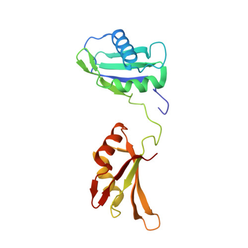
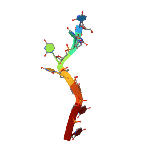
Structure-guided U2AF65 variant improves recognition and splicing of a defective pre-mRNA.

Agrawal, A.A.McLaughlin, K.J.Jenkins, J.L.Kielkopf, C.L.

(2014) Proc Natl Acad Sci U S A 111: 17420-17425

  • DOI: https://doi.org/10.1073/pnas.1412743111
  • Primary Citation of Related Structures:  
    4TU7, 4TU8, 4TU9

  • PubMed Abstract: 

    Purine interruptions of polypyrimidine (Py) tract splice site signals contribute to human genetic diseases. The essential splicing factor U2AF(65) normally recognizes a Py tract consensus sequence preceding the major class of 3' splice sites. We found that neurofibromatosis- or retinitis pigmentosa-causing mutations in the 5' regions of Py tracts severely reduce U2AF(65) affinity. Conversely, we identified a preferred binding site of U2AF(65) for purine substitutions in the 3' regions of Py tracts. Based on a comparison of new U2AF(65) structures bound to either A- or G-containing Py tracts with previously identified pyrimidine-containing structures, we expected to find that a D231V amino acid change in U2AF(65) would specify U over other nucleotides. We found that the crystal structure of the U2AF(65)-D231V variant confirms favorable packing between the engineered valine and a target uracil base. The D231V amino acid change restores U2AF(65) affinity for two mutated splice sites that cause human genetic diseases and successfully promotes splicing of a defective retinitis pigmentosa-causing transcript. We conclude that reduced U2AF(65) binding is a molecular consequence of disease-relevant mutations, and that a structure-guided U2AF(65) variant is capable of manipulating gene expression in eukaryotic cells.


  • Organizational Affiliation

    Center for RNA Biology and Department of Biochemistry and Biophysics, University of Rochester School of Medicine and Dentistry, Rochester, NY 14642.


Macromolecules

Find similar proteins by:  (by identity cutoff)  |  3D Structure
Entity ID: 1
MoleculeChains Sequence LengthOrganismDetailsImage
Splicing factor U2AF 65 kDa subunit
A, B
174Homo sapiensMutation(s): 1 
Gene Names: U2AF2U2AF65
UniProt & NIH Common Fund Data Resources
Find proteins for P26368 (Homo sapiens)
Explore P26368 
Go to UniProtKB:  P26368
PHAROS:  P26368
GTEx:  ENSG00000063244 
Entity Groups  
Sequence Clusters30% Identity50% Identity70% Identity90% Identity95% Identity100% Identity
UniProt GroupP26368
Sequence Annotations
Expand
  • Reference Sequence

Find similar nucleic acids by:  Sequence   |   3D Structure  

Entity ID: 2
MoleculeChains LengthOrganismImage
DNA (5'-D(*UP*UP*UP*UP*(BRU)P*UP*U)-3')C [auth P],
D [auth E]
7synthetic construct
Sequence Annotations
Expand
  • Reference Sequence
Small Molecules
Ligands 4 Unique
IDChains Name / Formula / InChI Key2D Diagram3D Interactions
CPQ
Query on CPQ

Download Ideal Coordinates CCD File 
O [auth B]N,N-BIS(3-D-GLUCONAMIDOPROPYL)DEOXYCHOLAMIDE
C42 H75 N3 O15
OJSUWTDDXLCUFR-HGZMBBKESA-N
SO4
Query on SO4

Download Ideal Coordinates CCD File 
G [auth A]
H [auth A]
I [auth A]
P [auth B]
Q [auth B]
G [auth A],
H [auth A],
I [auth A],
P [auth B],
Q [auth B],
R [auth B]
SULFATE ION
O4 S
QAOWNCQODCNURD-UHFFFAOYSA-L
GOL
Query on GOL

Download Ideal Coordinates CCD File 
J [auth A],
K [auth A],
L [auth A],
S [auth B]
GLYCEROL
C3 H8 O3
PEDCQBHIVMGVHV-UHFFFAOYSA-N
DIO
Query on DIO

Download Ideal Coordinates CCD File 
E [auth A],
F [auth A],
M [auth B],
N [auth B]
1,4-DIETHYLENE DIOXIDE
C4 H8 O2
RYHBNJHYFVUHQT-UHFFFAOYSA-N
Experimental Data & Validation

Experimental Data

  • Method: X-RAY DIFFRACTION
  • Resolution: 2.09 Å
  • R-Value Free: 0.223 
  • R-Value Work: 0.174 
  • R-Value Observed: 0.179 
  • Space Group: C 1 2 1
Unit Cell:
Length ( Å )Angle ( ˚ )
a = 164.432α = 90
b = 37.361β = 125.6
c = 100.192γ = 90
Software Package:
Software NamePurpose
SCALAdata scaling
PHENIXrefinement
PDB_EXTRACTdata extraction

Structure Validation

View Full Validation Report


Ligand Structure Quality Assessment 


Entry History 

Deposition Data

Revision History  (Full details and data files)

  • Version 1.0: 2014-11-26
    Type: Initial release
  • Version 1.1: 2014-12-10
    Changes: Database references
  • Version 1.2: 2014-12-24
    Changes: Database references
  • Version 1.3: 2017-11-22
    Changes: Database references, Derived calculations, Refinement description, Source and taxonomy
  • Version 1.4: 2023-12-27
    Changes: Data collection, Database references