


Structure of Mycobacterium tuberculosis protein
Korotkov, K.V.To be published.
Experimental Data Snapshot
Entity ID: 1 | |||||
|---|---|---|---|---|---|
| Molecule | Chains | Sequence Length | Organism | Details | Image |
| Probable nicotinate-nucleotide adenylyltransferase | 201 | Mycobacterium tuberculosis H37Rv | Mutation(s): 2 Gene Names: LH57_13225, nadD, P425_02519, Rv2421c, RVBD_2421c EC: 2.7.7.18 | 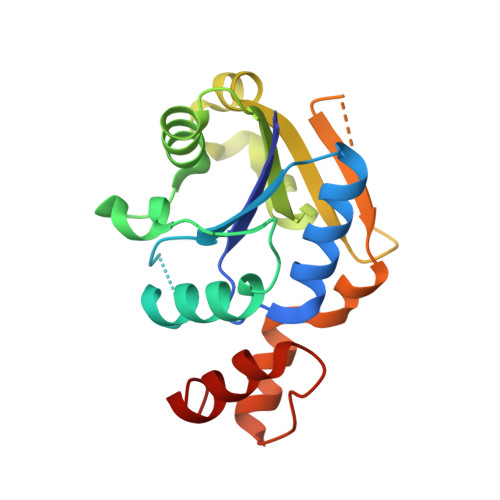 | |
UniProt | |||||
Find proteins for P9WJJ5 (Mycobacterium tuberculosis (strain ATCC 25618 / H37Rv)) Explore P9WJJ5 Go to UniProtKB: P9WJJ5 | |||||
Entity Groups | |||||
| Sequence Clusters | 30% Identity50% Identity70% Identity90% Identity95% Identity100% Identity | ||||
| UniProt Group | P9WJJ5 | ||||
Sequence AnnotationsExpand | |||||
| |||||
| Ligands 2 Unique | |||||
|---|---|---|---|---|---|
| ID | Chains | Name / Formula / InChI Key | 2D Diagram | 3D Interactions | |
| NAP Query on NAP | C [auth A], E [auth B] | NADP NICOTINAMIDE-ADENINE-DINUCLEOTIDE PHOSPHATE C21 H28 N7 O17 P3 XJLXINKUBYWONI-NNYOXOHSSA-N |  | ||
| CL Query on CL | D [auth A], F [auth B] | CHLORIDE ION Cl VEXZGXHMUGYJMC-UHFFFAOYSA-M |  | ||
| Length ( Å ) | Angle ( ˚ ) |
|---|---|
| a = 66.06 | α = 90 |
| b = 66.06 | β = 90 |
| c = 165.21 | γ = 120 |
| Software Name | Purpose |
|---|---|
| XSCALE | data scaling |
| PHASER | phasing |
| REFMAC | refinement |
| PDB_EXTRACT | data extraction |
| SERGUI | data collection |
| XDS | data reduction |
RCSB PDB (citation) is hosted by
RCSB PDB is a member of the

